Thursday 28 June 2012

Much a-flu about nothing revisited (post 2 of 2)

Welcome back to those who read yesterday’s post on the Kawaoka influenza paper. For those of you who have started reading this and haven’t read the other blog post, I’d suggest going back as we are halfway through a topic.

At the same time as Kawaoka’s team were working on their hybrid influenza virus in the USA another team lead by Ron Fouchier in the Netherlands were working on a similar project. The aim was again to look for mutations that would allow H5N1 to transmit effectively in a mammalian host. The big difference between the two studies is that Fouchier’s lab used a full blown H5N1 virus; as opposed to a hybrid. Fouchier’s paper was finally published last Friday in Science. Similarly to Kawaoka’s study, Fouchier began with an H5N1 virus isolated from a patient (in Indonesia). From this starting point the team added two mutations to the HA protein which are known to alter the specificity to α2,6 receptors (which, you may remember, is the first hurdle to allow mammalian spread). The mutations they added were Q222L and G224S. Some of the more astute readers may notice that this is our 
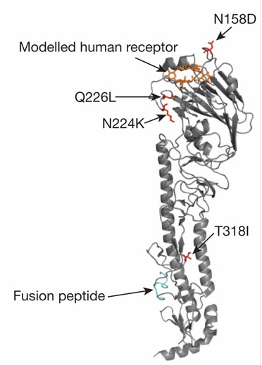
second encounter with position 224 of HA as the mutation N224K was seen in the Kawaoka paper. The fact that the position 224 of the virus from patients can either be a G or an N indicates how variable this virus can be. A third mutation was added in a protein called PB2 which forms part of the polymerase complex (a group of proteins needed to produce copies of the influenza genetic material). The mutation was E627K and allows the virus to replicate more effectively at the lower temperatures seen in the mammalian upper respiratory tract as opposed to the bird intestine (influenza is a gut infection in birds). The addition of these three mutations to the H5N1 virus did not allow for aerosol transmission between ferrets, even though the virus could bind to α2,6 receptors.
Cartoon of the influenza virus structure

Having failed to produce an H5N1 virus capable of aerosol transmission between ferrets with the three specific mutations, the team moved on to use the age old technique of passaging in order to force the virus’ evolution. Fouchier’s team took their triple mutant virus and inoculated a ferret intranasally (injected it to the ferret’s nose). After 4 days they would take virus from this ferret and do the same into a new ferret. After 10 ferrets had been infected in this manner the team produced viruses that were capable of aerosol transmission. Passaging is a beautiful example of how evolution works as only the viruses that have good replication are selected to grow in the new ferret, so replicative ability is selected for. From passage 7 to 10 the team induced sneezing in the ferrets to collect viruses that are best adapted for aerosol spread as well as replication. Thus this passaging process drives the evolution of viruses capable of a high level of replication in the upper respiratory tract and of aerosol transmission.

A coughing ferret
The team have therefore achieved their goal of making H5N1 viruses that are capable of aerosol spread, but that isn’t the end of the story. Fouchier’s team moved on to look at the additional mutations that had occurred to the triple mutant virus to allow aerosol transmission. They found that all the viruses capable of aerosol transmission had at least 9 mutations, including the initial 3. Interestingly, there were 5 mutations which were seen in all of the viruses capable of airborne spread; these being the 3 that were there initially along with T156A and H103Y of HA. The mutation of T156A is interesting as it comes very close to the N158D seen by Kawaoka and indeed causes the same effect of blocking a sugar binding to the protein. The H103Y mutation may play an important role in the stability of the HA protein similarly to the T318I of Kawaoka’s study.

Many press reports regarding the publication of the Fouchier study have claimed that only five mutations are needed for bird flu to become pandemic. I’d like to point out that that slightly misses the point. Five mutations are seen in all the viruses that became airborne in the study; however that does not mean these five mutations are sufficient for spread. It is likely that other mutations are also needed, hence the fact that the aerosol viruses all had at least nine mutations. What we can say is that between 5 and 9 mutations are needed as a minimum for aerosol spread of H5N1 in ferrets (I stress in ferrets as it comes back to the old point that ferrets are not humans). One other point that was sometimes missed in the press reporting of the paper was that none of the ferrets infected with the aerosol virus died, so similarly to Kawaoka’s study, there appears to be a loss of virulence when the virus becomes mammalian adapted.

Fouchier’s paper was published alongside a second influenza paper from Derek Smith’s lab at Cambridge (UK). This paper was looking for the presence of the mutations discovered by Kawaoka and Fouchier in the wild (both were co-authors on the paper). It was found that many H5N1 viruses are 3 and in some cases 2 mutations away from having Kawaoka’s 4 mutations and are 4 away from having the mutations cited by Fouchier. Again this became somewhat sensationalised in the press with headlines such as ‘bird flu is only two mutations away from pandemic.’ This drives me mad! If we turn those findings around they read somewhat differently; wild viruses have only 1 and in some cases 2 of the 4 mutations found by Kawaoka and only 1 of 5 mutations found by Fouchier. If we also add on the fact that these mutations are not necessarily sufficient for spread (remember Fouchier’s viruses had 5 core mutations, but all had at least 9) and the fact that they allow spread in ferrets, not necessarily humans, then it is starting to look less sensational. I’m not denying that bird flu has the potential to fairly easily mutate and become transmissible between humans, I’m just disappointed by the fear-mongering in some of the reporting.

Bird flu does have the potential to become a human pandemic, and pandemics are never good. I mentioned in my previous post that the current reported case fatality of bird flu is close to 60%. This number comes from the fact that there are very few confirmed cases of bird flu and of those that are known, 60% resulted in deaths. The thing is; flu is often a mild disease that people get better from in a couple of weeks so chances are many people won’t bother going to hospital and won’t be recorded as having bird flu unless it is serious, skewing the data (if it is serious enough for hospital then there is already an increased risk of death). Even if H5N1 is a highly virulent virus the Fouchier and Kawaoka studies both indicate that it loses virulence when it becomes airborne, so the idea that 60% of people who get it will die may be a long way off. Let’s not forget however, Spanish Flu of 1918 killed around 10-20% of those it infected, totalling 50-100 million deaths, so even if not at 60% bird flu is still a threat.

What needs to be taken from all this is that we now know the types of mutations needed for H5N1 to become mammalian transmissible. The two studies have shown that there are multiple routes for the virus to become airborne in the sense that they found different mutations; yet these mutations are seen to have similar characteristics. Changes are needed in the receptor binding site to allow α2,6 specificity. Mutations are needed to stabilise the protein to compensate for the apparent loss of stability seen when specificity is altered. It is likely that mutations are also needed in proteins other than just HA, for instance in PB2 (as in Fouchier’s paper). In order to effectively use this information it is essential to increase our understanding of the HA protein as this will allow even better surveillance for potentially risky mutations in the wild. While we continue to survey it is also essential that we work towards an H5N1 or universal flu vaccine and develop stockpiles of anti-viral drugs. That way, if a pandemic does start we are prepared and ready to respond as rapidly as possible. This was one of the major failings of the Swine Flu outbreak of 2009 in which people were caught off guard; hopefully we have learnt our lessons from that.

This blog was a bit more technical than some of my others so I hope I’ve managed to keep everyone interested. It is likely that this is not the last we will hear of bird flu so having a good understanding will help to avoid the fear-mongering which is so common in the press and allow you to assess the risk of a potential pandemic for yourself.

Wednesday 27 June 2012

Much a-flu about nothing revisited (post 1 of 2)

Last November I posted a blog regarding the biology of influenza virus and the controversial work of Ron Fouchier at the Erasmus Medical Centre in the Netherlands (here’s a link). A lot has happened since those blog posts. I’m sure many people have heard a little about the controversies surrounding the Fouchier paper and a similar paper by Yoshihiro Kawaoka, but I’ll give a brief summary for any that may have missed it. Two papers, studying the transmissibility of bird flu, were produced and submitted to the journals Science and Nature towards the end of last year. A huge debate then emerged over the fact that the studies contained potential dual-use. The argument was made that somebody may be able to use the information in the papers to weaponize bird flu. Since the virus has a reported case fatality rate of close to 60% (something that is in itself debatable) there is a large fear that the virus could be used for bio-terror attacks. As a result, the United States based National Science Advisory Board for Biosecurity (NSABB) voted in favour of blocking publication of the two papers. However, following certain revisions to the papers and clarification of some of the issues, the NSABB reversed its decision and voted in favour of publication in March this year. Both papers have now been published in their respective journals after an 8 month wait. Being that I posted about this issue back when it started, I thought it would be nice to do a follow up post looking at the two papers that have caused such a furore.

 Both of the papers are looking at the ability of avian influenza to transmit between mammalian hosts. Since it is generally frowned upon to use humans for such an experiment the researchers used ferrets which act as a good model to study how the virus could potentially spread in humans. Ferrets aren’t humans (in case there was any confusion), so even though they act as a good model for study, what holds true in ferrets does not necessarily hold true for humans. I don’t usually explicitly encourage people to read my other blog posts (I like to think that reading one may inspire you to look at more), however I am making an exception here since it may help with understanding of this post for anyone who is new to influenza. So here are the links to part 1 and part 2

Now that everyone is up to speed I’ll get into it. The first paper was published by Nature in May this year and came from the lab of Yoshihiro Kawaoka of the University of Wisconsin-Madison. As I described in my previous posts the key difference between avian influenza and a mammalian counterpart is in the receptor specificity. Avian influenza binds to receptors carrying an α2,3 linked sialic acid whereas mammalian viruses target α2,6 receptors. In order for H5N1 to transmit between humans the haemagglutinin (HA) protein must develop the ability to bind to α2,6 receptors. This was the starting point for Kawaoka’s team who added random mutations to a collection of H5N1 viruses isolated from a patient in Vietnam. The team then took all of their mutant viruses and selected those that were able to bind to α2,6 receptors. The H5 with the highest affinity to α2,6 receptors was found to have three mutations; N158D, N224K and Q226L. For the non-scientists reading, the letters refer to amino acids (the building blocks of proteins) and the numbers refer to the position of the amino acid in the HA protein. N158D means that the N (asparagine) amino acid at position 158 has mutated to a D (aspartic acid). The alterations at 224 and 226 are in the region of the protein which binds the receptor. The mutation at 158 prevents the addition of a sugar to the protein.

Having produced an H5N1 virus with specificity to α2,6 receptors the team moved on to produce a hybrid virus. The hybrid virus was made of the mutant H5 protein on an H1N1 virus backbone. In my previous blog I described the influenza genome as similar to a jigsaw with 8 pieces. Kawaoka’s team essentially took away a piece of the H1N1 jigsaw (the H1) and replaced it with a piece from the H5N1 jigsaw (the H5). This hybrid virus is therefore a full H1N1 virus, except for the replacement of H1 with H5. The H1N1 backbone is from the virus type responsible for the Swine Flu outbreak in 2009 and is still responsible for many flu cases now, meaning it is known to transmit in mammals. The only hindrance to the spread of this hybrid between mammals is therefore the HA protein taken from the mutant H5N1. The hybrid H5N1 was found to transmit through the air to two out of six ferrets. So while airborne transmission is occurring it is fairly ineffective.
Studying airborne transmission. The ferrets are separated by wire mesh so not to contact each other but can have spread of air between the two halves of the cage (and therefore hopefully spread of the virus).

The team then isolated virus from one of the infected ferrets, which brought to light an additional mutation. This mutation is T318I and was found to alter the stability of the HA protein. When the three initial mutations occur the protein becomes less stable which may explain the limited transmission. The T318I mutation compensates for the loss of stability and improves transmissibility. The virus containing all 4 mutations (N158D/N224K/Q226L/T318I) was much more effective at aerosol spread between the ferrets and at replicating within them than the triple mutant virus. However, even though the virus could spread easily between the ferrets, it was not seen to be lethal to them, indicating the virus loses some of its virulence in order to be transmissible.

The HA protein and locations of the mutations.
Image taken from the published paper (linked above)
So what does all that tell us? Kawaoka’s team have shown that four mutations to the H5 protein can allow it to effectively transmit between ferrets. With that knowledge it will be possible to survey H5N1 viruses in the wild and look for the presence of these mutations, thus allowing us to prepare for any potential pandemic. As I’ve already mentioned ferrets are not humans, so while these specific mutations have allowed spread in ferrets they may not necessarily allow spread in humans. Any surveillance should not be blinkered to these exact mutations, a fact which I will come back to when discussing the Fouchier paper. The results also hint at a loss of virulence when the virus can transmit by an aerosol route. This may occur if H5N1 naturally acquires the ability for aerosol spread in mammals, meaning the potential pandemic may not be as bad as feared (though this is mere speculation on my behalf and hard to test).

I don’t like to take up too much of people’s time with these blogs so look to keep them as short as possible. As such, I’m going to leave this post here. I’ll post the second part discussing the Fouchier paper tomorrow to round it all off. So you’ll have to come back to find out about that one.

Wednesday 13 June 2012

Caspase: the not so friendly protein

It’s somewhat of an antithesis to say that death is essential for life, however in multicellular organisms, such as humans, without cells dying we would not live. The topic of this week’s blog is therefore a fundamental phenomenon of all cells in our body termed apoptosis, or programmed cell death. As the name implies, this form of cell death is highly controlled and essentially runs a program to cause the death of the cell. Apoptosis is controlled by a set of proteins called caspases which drive the necessary changes to kill the cell.

Hand sculpting
So why do cells need to die? There are a lot of answers to that question but I will only look at a few of these. Firstly, right back when we are a foetus we all start with webbed hands and toes (see the image). However, I assume most people no longer have such webbing, a fact which all comes down to death of the excess cells between the digits. Another example of this sculpting process can be seen when a tadpole matures to a frog (the tail isn’t chopped off by a small frog knife).

Human embryo hand at 8 weeks

Two further roles for apoptosis can be seen during infection. Viruses enter cells and use them to produce many new viruses, causing damage to the host. One of the front line defences against this is to simply kill the infected cell, which blocks replication of the virus. Also when we are infected with any pathogen an immune response is produced. This response is controlled by white blood cells, our army against the invading infectious organism. When an organism enters the number of white blood cells rapidly increases, in a sense, the army begins conscription. When the infecting organism has been removed from the body all these excess cells must be removed otherwise they could cause autoimmune disease (where they attack healthy tissue causing disease), or could continue growing in number, causing a cancer. This removal all comes down to cell death.

The final example for the importance of apoptosis I would like to look at is in development of the brain. Our brains are initially made with a huge excess of neurones. Interesting these cells (like many others) are naturally programed to die; death is the default setting. The cells in the brain only survive if they are able to make contacts to other cells (forming synapses). The cells that connect in the brain survive and produce the brain of the foetus; those that make no contacts undergo their default setting and die, so as not to clog up the brain with excess, useless neurones. Around 50% of the initially produced neurones will die because of not making any contacts. Apoptosis is therefore an essential function for life, without it we would not progress beyond being an embryo.

So now the question to ask is what causes cells to die. As I mentioned, the key players of cell death are proteins called caspases. Caspases are expressed in all cells as zymogens, a fancy word for an inactive enzyme. These zymogens sit in the cell waiting for any time there is a trigger to activate them. This can be thought of almost like a light switch, everything is there waiting to be turned on, but it needs you to press the switch to get light. In the cell there are two main pathways that lead to activation of the caspases; intrinsic and extrinsic pathways. Intrinsic signalling relies on proteins in the cell which form pores in the mitochondria (the molecular machine that produces energy in our cells). These pores cause the release of two proteins, called cytochrome C and Apaf1, which together bind a certain caspase (caspase-9) and cause its activation in the ‘apoptosome.’ The extrinsic pathway relies on a receptor on the surface of the cell binding a signal (this is the case in the immune response for killing infected cells.) When the receptor engages with its target protein a platform is set up inside the cell called DISC (death inducing signalling complex) which binds capsase-8 and activates it. While both pathways activate different caspases the effect is the same; RIP cell.

Apoptotic cell blebs being engulfed by a macrophage
With the activation of caspases our poor cell is terminal. Many changes occur to the cell and these are all controlled by the activity of caspases. Firstly the cell begins to shrink as all the proteins that keep a cell at its usual size are broken up. Over time the DNA inside the cell is chopped up into tiny fragments, rendering the cell useless. Eventually parts of the cell start breaking off in small units called blebs, until at the end a big macrophage (an immune cell) comes along and eats up the cell. This may sound like a pretty nasty way to go, shrinking down, having the DNA chopped up and then being eaten by a giant, but this process protects the rest of the organism from damage. Other forms of cell death often involve the cell bursting which causes inflammation in the surrounding area and therefore more extensive damage. Apoptosis, on the other hand, is completely silent therefore providing the most effective and safest way to kill a cell.

It fascinates me how essential cell death is for life. Without death we could not be born; it’s that fundamental. We continue to need cell death right up to our own death as it can protect from cancer, aid in clearance of infection and may help to stop autoimmunity developing. However too much death is obviously not a good thing either and is seen in many neurological disorders, so it’s a fine balancing act between the good and the bad of cell death. It’s also interesting to think of cell death in an evolutionary sense. When life began it was only as a single, isolated cell whose only purpose was to survive. As life became more complex and organisms made of multiple cells emerged, the ability to kill off a few cells to protect the organism as a whole became a huge advantage. Perhaps the evolution of this self-sacrifice was an essential step in the development of complex life itself…

Thursday 7 June 2012

There’s a legion of bacteria out there, but one is stealing the headlines

This week, the UK headlines (the Queen’s Diamond Jubilee aside), have been dominated by an outbreak of Legionnaires’ disease in Edinburgh. It therefore seems fitting that I should join the trend and write my first blog post in a fairly long time on this topic.

Legionnaires’ disease first acquired its name back in 1976 when a Philadelphia based convention of the American Legion was struck by a plague of pneumonia. It took almost 3 years for the causative agent to be discovered, which was traced to a cooling tower containing the bacterium named Legionella pneumophila. The Legionellae bacterial family is large, with roughly 50 different species, yet only a few of these have been found to cause any kind of disease. Legionellae bacteria are classed as being “Gram negative” meaning they do not take up a marker stain for bacteria known as the Gram stain. Gram negative bacteria are characterised by a large protective layer surrounding their cell known as the outer membrane. This outer membrane can help to protect the bacteria from certain antibiotics and thus limit the efficacy of certain treatments, such as penicillin. Legionnaires’ disease outbreaks are sporadic events and are often traced to man-made structures like cooling towers and air conditioning units. The outbreaks are characterised by pneumonia and flu-like symptoms. Those who have read previous blogs of mine may remember the discovery of Mimivirus, which was originally thought to be causing a Legionnaires’ disease outbreak (here’s a link to refresh the memory or for those who haven’t read it before.)

L. pneumophila are predominantly found in freshwater environments. When this was first discovered it seemed odd since growing L. pneumophila in a lab was very difficult due to the need for a very high level of nutrients, which are not naturally found in freshwater. It became apparent that these bacteria do not grow in a completely isolated way; much like a virus, the bacterium needs to parasitize another living organism; in the case of L. pneumophila the main host species are amoeba.
Interestingly, most natural freshwater reservoirs tend to normalise to the ambient temperature, so a collection of rain water inside an old tyre, for instance, would be at the temperature of the surrounding air (I use the tyre example as I will soon be writing a blog on Dengue virus - you’ll have to come back to see why tyres are important). However, L. pneumophila grows best when at 25 OC - 42OC, with the best growth occurring at 35 OC. Now I haven’t been to Scotland in a long time, but I’m pretty sure it hasn’t got up to 35OC any time in the recent past (if ever). While natural freshwater reservoirs are unlikely to reach high enough temperature to support L. pneumophila growth, man-made environments such as cooling towers or air conditioning units are. Legionnaires’ disease is therefore, in essence, a man-made phenomenon due to our alteration of the environment (it’s not alone in this respect).
So the next question to look at is how exactly this little bacterium is causing the outbreak of the disease we are currently seeing in Scotland. There are two main ways L. pneumophila can jump from its usual amoeba hosts into humans; these can be either through small droplets of water containing bacteria that are inhaled into the lungs or through an aspiration route in which water in the mouth containing bacteria gets into the lungs. The aspiration route needs some pre-existing damage as things in the mouth are not usually meant to enter the lungs; this damage can be caused by smoking or chronic lung disease for instance. Our lungs are lined with immune cells known as phagocytes which essentially sit there and wait for things that shouldn’t be there, such as bacteria. Once the phagocytes detect the bacteria, they engulf then and taken them into the cell where they would normally be degraded (a process known as phagocytosis), thus providing protection. L. pneumophila are a bit cheeky. The bacterium allows itself to be phagocytosed but then blocks the destruction step; they simply remain in a compartment of the cell where they are able to grow. Over time the growth of L. pneumophila will lead to death of the infected phagocyte which is often accompanied by bursting of the cell. This bursting, like popping a water-balloon, releases all the contents to the environment. All the bacteria and all the other nasty stuff inside a cell are released out into the lungs causing a large inflammatory response. It is this response, designed to protect, that causes the flu-like symptoms and the pneumonia seen in Legionnaires’ disease. When this becomes severe (which is relatively rare) people need hospitalisation for treatment. The treatment is generally effective, so for most people Legionnaires’ is a nasty disease but one that usually clears up by itself or through medical intervention.
As I finish writing this post, there are currently 24 confirmed cases of Legionnaires’ disease in the current outbreak, with one death. It’s a nasty disease but one that is rare and can very often be treated and I’m sure that within a few days to a week the number of new cases will begin to decrease.