Tuesday, 1 March 2016

Fighting cancer with a virus of plants: it's not a load of bull


If I ask you to think of a virus, I’m going to guess you’ll land on one of Zika, Ebola, HIV, Influenza or maybe at a push, Rhinovirus, the latter two of which you may be unfortunate enough to have recently experienced in the winter of the northern hemisphere. Viruses, and the infections they cause, are massive public health concerns, and for the most part this is what gets our attention. However, there is currently a vast amount of research going into the development of viruses as tools in the fight against cancer, and it’s some of this work I’d like to discuss in the blog post.

The work I’m going to talk about was published in the journal Nature Nanotechnology and comes from the Geisel School of Medicine in New Hampshire, USA. The published article has the somewhat striking title of “in situ vaccination with cowpea mosaic virus nanoparticles suppresses metastatic cancer.”

Let’s break that down. Our immune system has the capacity to destroy cancer cells, this is in fact one of the major hurdles a mutant cell needs to overcome in order to develop into a cancer. In order to escape the immune response, mutated cells need to develop ways in which to protect themselves, for instance, by releasing molecules that act to suppress the the immune system - in a sense, camouflaging themselves. This kind of immunosuppression by cells is nothing strange in our body, it’s part of a normal response to stop your immune system attacking your own body. However, cancer cells develop the ability, through mutation, to subvert this system to aid their own growth. As such, an idea that has starting to come to the front of cancer research is so called cancer immunotherapy; stimulating the immune response to fight against cancerous tissue, while leaving normal tissue untouched. So to break down the first part of the paper’s title, “in situ vaccination” is the idea of directly supplying something to trigger an immune response at the site of a cancer. The “something” that is being supplied into cancerous tissue in the case of this work, is cowpea mosaic virus nanoparticles.  
 
Cowpea peas
 
I’m guessing that cowpea mosaic virus isn’t something you’ve come across before (I certainly hadn’t). By way of background, cowpea mosaic virus, is a virus that infects the cowpea plant, and causes a mosaic pattern on the leaves (to state something you’d probably guessed). In this study of cowpea mosaic virus as an anti-cancer agent, the group work with “nanoparticles," which can also be called "virus-like particles." If you consider a virus particle, such as the one depicted for cowpea mosaic virus (below), that 3D shape is made up by very few proteins. A virus structure is essentially just a protein case for the genetic material. A virus-like particle is simply just this casing with nothing inside (literally a shell of it’s former self). Without the genetic material, these particles are no longer able to infect cells. An analogy for this could be a laptop that has had all of the internal circuit boards removed. From the outside it still looks like a laptop, but try and turn it on, and nothing will happen. Virus-like particles are of very little danger because they cannot infect cells, however, the immune system isn’t able to detect this. The cells of the immune system only ever see the outer casing of a virus and treat this as a threat - thus virus-like particles can stimulate an immune response just like a real virus (from the outside a laptop without circuit boards still looks like a laptop). And this is the basis of some vaccines, most notably of which is the vaccine against human papillomaviruses, which some readers may have received. 
 
Cowpea mosaic virus structure (from Wikipedia)
 

We can now get to the meat of the paper. To jump to the punchline, the group were able to show that injecting mice with cowpea mosaic virus-like particles could stimulate an immune response, that resulted in suppression of cancers in mice. Initially the group determined that treating cells with the virus-like particles was capable of triggering an immune response in a dish, and moreover, found that this treatment didn’t cause the cells to die. The group moved on to have mice inhale preparations of cowpea mosaic virus-like particles. In mice with no tumours, this inhalation resulted in activation of the immune system, which could be detected by an increase in the number of neutrophils (one class of immune cells) in the lungs. And indeed, collecting neutrophils from these mice showed that the virus-like particles had been taken up into these cells, thus activating them - the neutrophils treated the virus-like particles as a true threat and attempted to remove them. Importantly, this inhalation of the virus-like particles to healthy mice didn’t cause any adverse side effects or damage to the lungs.

The next experiment was to give the virus preparation to mice with tumours. It was found that before the inhalation, the immune cells in the lungs of these mice was very different to that of healthy mice. There were far more cells associated with immunosuppression (as a result of the cancer). Upon giving virus-like particles by inhalation to these mice, it was seen that there was a large increase in the number of activated immune cells capable of tackling cancer (again, a large increase in the number of neutrophils, for instance). So far then, everything seems to be going to plan for using cowpea mosaic virus to activate an anti-cancer immune response.

Next, the group started to give mice injections of the virus-like particles instead of allowing them to inhale. They found that injecting mice with cowpea mosaic virus-like particles resulted in a decreased number of tumours in the lungs. Importantly, the researchers nicely demonstrated that this reduction in tumour number was due to an immune response but using mutant mice that don’t have a functional immune system - these mice showed no change in their tumour burden.

The tumours that were being tested to this point were melanomas growing in the lungs. But it wasn’t just melanomas that showed sensitivity to the immune response triggered by cowpea mosaic virus-like particles. In one set of experiments, mice were injected with cancerous tissue in their fat pads, which then spontaneously metastasis to the lungs 16 days later. The mice given injection of virus-like particles all showed delayed onset of lung tumour formation, and survived the treatement. Similarly, mice with tumours in their colons could also be protected by treatment with the virus-like particles. 

Finally, the group tested whether injections of virus-like particles could protect against melanomas growing in the skin. Once again, they found that cowpea mosaic virus-like particles could protect the mice, and half of the mice injected completely cleared their tumours after only two injections of virus preparations. Moreover, this anti-tumour immune response was found to be long lasting, since when mice that had previously cleared tumours were injected with more cancerous tissue at a later date, 3 out of 4 completely rejected the implanted cancer (while this cancer grew in all non-vaccinated mice). 

Overall this work has nicely demonstrated the potential for the use of cowpea mosaic virus-like particles in the battle against cancer. These virus-like particles could be tolerated in mice and were seen to trigger an immune response, which resulted in clearance of cancerous tissue. The virus-like particles showed no major side effects, and even seemed to provide long lasting immunity against the cancers used in the work. The next step for cowpea mosaic virus will probably be to test safety in humans. It will take a long time, and many more studies, but perhaps a virus that infects plants could one day be used to treat cancers in humans.

Sunday, 31 January 2016

From nowhere to everywhere - Zika virus


It seems to have come out of nowhere, and spread like wildfire. A new virus is rampaging through Brazil and parts of the Americas, and causing major global concern. Just as Ebola finally subsided in Africa, enter Zika virus from left stage. I’ll be honest, until a couple of weeks ago, I’d never heard of Zika virus, and I’m probably not alone. So I’ve decided to put together this blog post to discuss some aspects of this virus and the current outbreak. 
 
Countries where Zika virus has been found - from the New York Times
 

To being, let’s look at the virus itself: Zika virus is a member of the Flaviviridae family, which includes the much better known dengue, West Nile and yellow fever viruses, and like these is transmitted by mosquitoes. Because no-one really cared about Zika until recently, the virus has little published scientific literature. Typing "Zika virus" into PubMed (the major search engine of academic literature) yields 155 published articles, compared to the 9198 that are returned from searching "dengue virus” (at the time of posting). However, being that we know the virus family, this already tells us a lot. Like other flaviviruses, Zika is small with a genome of just 10.8 kilobases of positive sense single stranded RNA, which can encode 10 genes. The genome being positive sense and single stranded means that once it is released into a cell, the RNA will be treated just like any other cellular, protein encoding, RNA and interact with ribosomes to produce new protein. The 10 proteins that are produced by a cell following a Zika virus infection are responsible for taking over that cell and replicating the virus so it can spread new particles to new cells. Again, as with other flaviviruses, the Zika particle is small, and has one protein, the E protein, protruding on the surface. This protein is involved with attaching to a cell and responsible for the subsequent events that culminate in release of the genome into that cell. The E protein also makes up the main target for the immune system to tackle the virus. 
 
The structure of a flavivirus - from ViralZone
 

Zika virus was first identified in 1947 from a Rhesus macaque monkey which was being used as sentinel for yellow fever. At the time, sentinel monkeys were commonly used to detect the presence of yellow fever in an area since tests for viruses weren’t quite what they are today (at the time there weren’t even cell line to use in a lab to infect with the virus to study it!). This particular monkey, Rhesus 766, developed signs of a viral infection while in the Zika forest of Uganda. Blood was taken and used to inoculate mice, all of which became sick. Virus was isolated from the brains of deceased mice, but determined to be different from yellow fever - thus Zika virus was defined. The first human cases of the viral infection were reported in 1968 from Nigeria. Zika virus then remained largely undetected outside of Africa and parts of Asia, until 2007 and 2013 which saw epidemics in Yap Island and French Polynesia, respectively. 

Zika virus generally causes very mild symptoms in human infection, if any symptoms at all - only about 25% of cases are symptomatic. For that reason, little attention had been paid to it, until recently of course. The 2013 outbreak in French Polynesia was the first real indication that Zika infection could have more serious consequences than just rash, fever, malaise, and all the other general symptoms the initial infection can cause. During the outbreak, a spike in the cases of Guillain–Barré syndrome (GBS) were reported, with 73 individuals being diagnosed with the disease. GBS results from damage to the perisperhal nervous system (i.e. not the brain or spinal cord), causing rapid onset muscle weakness, and in severe cases, can result in paralysis. GBS can be life threatening since the peripheral nervous system is responsible for controlling the muscles involved with breathing and the heart beat. This coincidence of Zika virus spread and GBS was the first indication that Zika could potentially damage the nervous system.

So that brings us to the current outbreak occurring in Brazil, where once again, there appears to be circumstantial evidence linking Zika virus to damage of the nervous system - this time in the form of microcephaly in new born infants. Microcephaly, meaning abnormal smallness of the head, can be caused by multiple factors. Other infections can be responsible, such as rubella virus, cytomegalovirus or toxoplasmosis. As can poisoning of a foetus from alcohol, mercury or radiation. Mother malnourishment and diabetes may also be linked to microcephaly. In about 15% of microcephaly cases, the infant just has a head of smaller size, but in other cases, this small head is connected with poor development of the brain which can result in developmental delays, intelligence deficits and hearing loss, as well as premature death.  
 
Microcephaly - from Wikipedia
 

In the previous paragraph you may have noticed my wording that there is circumstantial evidence linking Zika virus to microcephaly cases; it is not clear that Zika infection is responsible. However, as Zika has begun to spread in Brazil and parts of the Americas, there has been a phenomenal spike in the number of microcephaly cases. Until 2014, there were around 150-200 cases of microcephaly in Brazil each year. The birth index for Brazil is estimated at 14.72/1000 population, in a country of around 200 million, this means just under 3 million as an estimate for the number of births in Brazil each year - so cases of microcephaly were rare. However, in 2015, there were nearly 3000 microcephaly cases, with the vast majority of these being reported in the latter part of the year, right around the time Zika virus cases started to become more common.

As I’ve stated, it is still not clear that Zika virus is responsible for the rise in microcephaly cases, though it is clearly the major culprit. More time is needed to follow pregnant women and determine if they become infected with the virus during their pregnancy. However, there will still be difficulties to determine if Zika alone is responsible. The best way to confirm a Zika virus infection is to look for the viral RNA genome in the blood of patients, however, the time in which this can be done is limited to the first week of infection. After that, diagnosis relies on finding antibodies against the virus in a patient’s blood. However, these serological tests are complicated by the fact that antibodies against Zika virus can cross-react with dengue virus - making it difficult to tell which virus was truly responsible for the presence of those antibodies. That dengue virus is endemic in Brazil, and the vast majority of people are positive for antibodies against dengue, compounding the issues with truly detecting Zika virus. However, with studies now aimed at determining if Zika virus is truly responsible for microcephaly, pregnant women will be tracked much more carefully, therefore raising the prospects of being able to catch an infection in the first week where viral RNA can be used to confirm a Zika virus infection.

Finally, what can be done? The short answer is not a great deal. We currently have no antiviral drugs to tackle any flavivirus infection, let alone Zika. Work is under way around the world trying to find broad-spectrum antiviral drugs that could be used to combat infection from multiple viruses, but whether a drug intervention would be of much use in a disease that generally causes few symptoms, except in pregnant women who may be unable to take the drugs, remains unclear. A better prospect would be the development of a vaccine, but we are a long way from that. Vaccines to Ebola were starting to undergo trials towards the end of the recent West Africa outbreak, but these had been years in the making through research on Ebola. Very little study has gone into Zika virus and potential for vaccines against it. However, there is a vaccine against yellow fever virus, and one idea would be to modify this to have the E protein of Zika, instead of yellow fever. The yellow fever vaccine is one of the best vaccines we have developed, but generating a Zika vaccine, and testing it, will take many years. Making things worse is the ethical issues of testing the vaccine in the population that seem most at risk from Zika virus, pregnant women. 


For the time being, our best option may be to tackle the mosquitoes which transmit the virus. There are the basic measures of wearing clothes to cover the skin and the use of bed nets when sleeping. Additionally, clearing standing water pools, such as in flower pots, or the inside of old tires, where mosquitoes lay their eggs will help to disrupt the mosquito life cycle. And in terms of more complex measures, many tests are under way to release genetically altered male mosquitos which cannot produce viable offspring. Additionally, work on the bacteria Wolbaccia may still hold hope for reducing the risk posed by mosquitoes (this is something I’ve previously posted on). Tackling mosquitoes seems to be one of the biggest challenges we face in the coming years. The Aedes species, responsible for transmitting Zika virus also transmit dengue virus and Chikungunya virus. The Culex species transmit West Nile virus, and the Anopheles species transmit malaria. Along with global warming, the habitable zones of these mosquitoes will spread, putting more people at risk. The best way we can tackle the dangers posed by mosquitoes is through continued scientific research, both on the mosquitoes themselves, and the viruses they harbour.

The most pressing issue now for Zika virus is determining that it is indeed the virus that is causing microcephaly, and finding ways to tackle its spread by mosquitoes. 

Monday, 20 July 2015

Hate mosquitoes, this work will help you avoid them


My two most recent posts to this blog have had an, admitted unplanned, theme of looking at viruses spread by Aedes mosquitos (Dengue and Chikungunya). Shortly after posting my last article, research was published which analysed the global distribution of the two Aedes species responsible for spread of these, and other viruses. So it seemed like a logical way to conclude this mini-series of posts. (I highly recommend going to the hyperlink for the paper as there are some cool maps to look at at the bottom).

Aedes mosquitoes are limited to where they can live by multiple factors, predominantly temperature and rainfall. The insects need high temperatures, but also need rainfall as they lay their eggs in water. These factors help to limit the habitable zone of Aedes largely to the topics. However, with global warning, the regions in which the mosquitoes can survive and spread is increasing, causing concern about the viral diseases they can transmit.

The study, which was published in the journal eLife, aimed to establish models to predict the global regions that Aedes egypti and albopictus could live. The models were based on real world data gathered by capturing mosquitoes across the globe. In total, 19,930 A. egypti and 22,137 A. albopictus mosquitoes were captured. Most of these were captured in Asia, 60% of the A. egypti and 75% of the A. albopictus captures. The Americas came in a comfortable second for the highest proportion of captures. This suggests that most of the mosquitoes currently reside in tropical parts of Asia and the Americas. Building on this knowledge, and factoring in climactic conditions that impact mosquito survival, models were generated to predict potential areas that the mosquitos can live.

Using these models, we now have more information about areas that could be at risk of the viral diseases carried by the mosquitoes. For instance, Mexico is predicted to have ideal conditions for the two mosquito species, even though none were captured there during the course of the study. In Europe, captures suggested that Greece and Italy have an increasing presence of the mosquitoes, and that while none were captured there, Spain, Portugal and much of south-eastern Europe have good conditions for spread.
A large part of this work is predictive, but it is based on a lot of data both on capture of mosquitos and climactic conditions. The knowledge that has been generated about current regions in which the mosquitoes spread, and the regions with potential, will direct public health work. Keeping an eye on whether the mosquitoes become established in Spain for instance, will help people take better precautions and look to limit the spread of viral diseases such as Dengue and Chikungunya. Forewarned is forearmed as they say.

Thursday, 25 June 2015

Chikungunya - don't be scared, but do know about it

Chikungunya virus and it's beautiful symmetry
In my previous blog post I discussed advancements towards a vaccine against Dengue virus. I’ve decided to keep with the topic of viral infections that, along with global warming, are now spreading out of the tropics and causing concern. The topic this time is Chikungunya virus. My hope for this post is to tell you a little about a virus you’ve probably never hear of, but if you take nothing else from it, you’ll at least be able to pronounce the name - chihk-uhn-guhn-yuh.

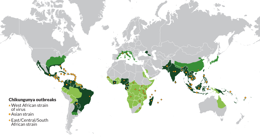
The habitable zone in which mosquitoes can survive is spreading because of global warming, and this is starting to pose some serious public health issues. Chikungunya was first described in 1952, and until recently, has been predominantly confined to Africa and parts of Asia. Yet as Aedes mosquitoes, the vector for the virus, spread, so too does the disease, which has now been found in the Americas and even parts of Southern Europe. Estimates suggest there have been over 3 million cases globally.

The virus is typically carried by the Aedes aegypti mosquito whose geographical range is restricted to the tropics. However, in 2005, a new strain of Chikungunya was found with a single mutation to one of the proteins exposed on the surface of the virus that allowed it to replicate efficiently in the closely related Aedes albopictus mosquito species. This mutation, and change of vector, was responsible for an outbreak on the island of Réunion, in the Indian Ocean, that caused around 266,000 cases (in a population of 800,000). A. albopictus can survive in more temperate conditions that A. aegypti, meaning this stain of the virus poses a real thread of widespread transmission. Indeed, this mutation is likely responsible for the emergence of cases in Southern Europe.


Global outbreaks of Chikungunya (https://www.sciencenews.org/article/chikungunya-move)

More recently Chikungunya has made its way to the Caribbean and the Americas, causing nearly 800,000 cases of disease across Caribbean islands last year. Fortunately, the virus that was transmitted across the Atlantic was not capable of spread in A. albopictus, which probably played a big part in limiting spread of infections to Florida and no further into the USA. However, the fact the virus is circulating causes concern; should mutations occur allowing spread in A. albopictus, much of North and South America will be at huge risk from endemic Chikungunya spread.

Chikungunya rarely causes fatal disease (probably explaining why not many people have heard of it), however, there are no current treatment options, nor a vaccine. While the infection may not be life threatening, that isn’t to say it’s pleasant. The disease begins like may viral diseases, causing the “flu-like” symptoms of fever, headache, chills etc. However, Chikungunya can also cause severe, even debilitating join pain, which can persist for months to years. Indeed, the name derives from a word in the Kimakonde language of East Africa meaning “to be contorted,” in reference to the appearance of infected individuals. Such symptoms can put real strain on working communities, and there are reports of whole towns coming to a stand-still because of bed-ridden Chikungunya sufferers.

Chikungunya is a virus that is currently sat on the brink of major global spread, yet I’m guessing most people have never heard of it. Should the virus currently found in central parts of the Americas mutate to spread in A. albopictus, a huge number of people will be at risk of a severely debilitating disease. It does seem that infection with Chikungunya can provide protection from re-infection, making the prospects of a vaccine promising. However, as was highlighted with the recent outbreak of Ebola, without proper forward planning, and funding, science is largely reactive; should Chikungunya spread really take off, I suspect we will not be ready for it. Even though it may not make the headlines as much as other viruses, research into understanding Chikungunya is necessary. Hopefully this blog has helped illustrate why it’s important to work on viruses other than those that always hit the headlines.

Tuesday, 9 June 2015

Maybe one less thing to worry about from a mosquito bite - prospects of a Dengue vaccine

Under the assumption that a majority of the readers of this blog are from the UK, I’m guessing not many have experienced, or even come across Dengue fever, or “breakbone fever” as it is often referred to. Any readers from the USA, central America, Australia, or other parts of the tropics are potentially acutely aware of the disease. The illness is characterised by high fever, malaise and bone/join/muscle pain which can be crippling (hence the colloquial name of breakbone fever). In extreme cases, Dengue can present with hemorrhagic fever (similar to the symptoms of Ebola).


Aedes mosquito


Dengue fever is caused by Dengue virus (DENV for ease), which is transmitted by Aedes mosquitoes. As with other mosquito borne diseases, due to global warming, the cases of Dengue fever are increasing because the habitable zone of the mosquitoes is expanding. Currently it is believed that 2.5 billion people are at risk of DENV infection. Currently, there are no approved drugs to directly tackle DENV, nor is there a licensed vaccine, even with intense research in the area. However, recent trials of a potential vaccine, produced by Sanofi-Pasteur have been very promising, and it is this which I will discuss here. I’d first like to tell you a little bit about DENV to explain why producing a vaccine has been so challenging, before looking at the results of the recent clinical trials and the potential for a licensed Dengue vaccine.

An image from http://www.healthmap.org/dengue/en/ of the cases of DENV in that last 3 months. Areas closer to red being those with most cases and most confidence.

DENV is a very interesting virus because of its interplay with the immune system. Let us use the example of measles virus to explain what I mean. When measles virus infects an individual (or they receive the vaccine), the immune response is activated and will produce antibodies specific to that virus. These antibodies bind to the virus and block it from infecting cells and promoting its destruction in immune cells such as macrophage, stopping the infection and protecting you. Those antibodies will do nothing against any other virus, but are very effective against measles.

An antibody response is highly beneficial against certain viruses, such as measles, but the situation is a little more complex for DENV. There are four very distinct types of DENV, which are known as serotypes, DENV-1, -2, -3 and -4. Should an individual become infected by DENV-1, for instance, they will produce protective antibodies against this serotype of the virus. If someone else is infected by DENV-2, they will produce antibodies specific to that virus. Antibodies specific to each serotype are only effective for that particular virus; DENV-2 antibodies will have no protective effect on DENV-1. This production of these specific antibodies is largely what dictates there being four DENV serotypes.

If an individual is infected with DENV-1, they will produce protective antibodies against re-infection by DENV-1. However, if that person should become infected by any of the other DENV serotypes (let’s use DENV-2), this can produce a life threatening situation. The antibodies specific to DENV-1 from the primary infection will bind to DENV-2, but instead of stopping the virus from infecting cells, the antibodies will promote infection of cells that are not normally infected by DENV - a phenomenon known as antibody dependent enhancement (ADE).
A diagram to depict ADE (from http://www.the-scientist.com/?articles.view/articleNo/34586/title/Antibody-Dependent-Enhanced--ADE--Immunity/). 1) Virus infects cells. 2) The immune response is activated and antibodies produced. 3) These antibodies bind to the virus and can result in uptake to, and 4) destruction in macrophage cells. 5) Upon a secondary infection from a different serotype, 6) antibodies are release from memory B cells. 7) These release antibodies do not bind the new serotype as effectively. 8) The virus can infect more cells, such as macrophages, leading to more extensive infection.
One such example of cells more susceptible to a secondary infection are macrophage cells. Under normal circumstances, a macrophage will detect virus particles bound by antibodies and destroy them. However, in the case of DENV and ADE, the macrophage are unable to destroy the different serotype of the virus, and will instead become infected. Once infected, these cells will eventually be killed, but can also spread the virus to the rest of the body, and even into the brain. This secondary DENV infection can cause much more extensive infection, and as a result, much more severe disease, often characterised by hemorrhagic fever or shock syndrome which can be life threatening.

The case fatality for DENV is only 1-5%, yet with around 50 million infections each year, and 2.5 billion people living in areas at risk from the transmitting mosquitoes, this is still a lot of people. It is speculated that of the fatalities, a large proportion come from a secondary infection.

A vaccine is generally designed to elicit an antibody response. In short, this has been why producing a DENV vaccine has been so difficult. If the vaccine only produces an antibody response against one serotype of the virus (let’s say DENV-1), that patient is then at more risk of having a severe DENV infection than they would be without the vaccine. In order for an effective vaccine against DENV to be produced, an antibody response against all four serotypes of the virus must be produced at once, so as not to leave the individual susceptible to more severe infection. This has proved very difficult.

There have been many failed attempts at producing a DENV vaccine. However, finally it seems like there may be light at the end of the tunnel. As touched on above, the vaccine has been manufactured by Sanofi-Pasteur and it is based on the yellow fever vaccine. The first vaccine ever produced was against smallpox, credited to Edward Jenner. Once the idea of vaccination became established, the next virus on the hit list was yellow fever virus. The vaccine against yellow fever virus is still one of the most effective ever produced. Yellow fever virus and DENV are from the same virus family (the Flaviviridae), and therefore share many features. The work at Sanofi-Pasteur used the yellow fever vaccine as a backbone for their DENV vaccine (known as CYD-TDV). The yellow fever vaccine was altered to present the immune system with proteins against each of the four DENV serotypes instead of yellow fever virus, then made as a cocktail of all four (a tetravalent vaccine).

The vaccine has so far been trialled on around 31,000 children (who are at most risk of severe DENV) in South Asia and Central America and in both regions has been show to be very effective. In the South Asian trial the vaccine had 56.6% efficacy for all DENV infections, and 80.8% efficacy at preventing severe DENV infections, and importantly, showed no substantial safety concerns. While in the Central American trial these equivalent figures stood at 60.8% and 80.3%. While efficacy of around 60% may seem low, this is still a vast improvement from no protection, but most importantly, the vaccine seems to be able to reduce the number of hospitalisations from severe DENV infection by up to 80%.

The vaccine is still yet to be licensed, but with the results from these phase III clinical trials, the prospects look good. Sanofi-Pasteur has already spent 20 years and $1.7 billion in the attempts to bring this drug to market, so not least for them, but also for the 2.5 billion people at risk of DENV infection, the prospects of a DENV vaccine are highly exciting. Hopefully later this year we will see the vaccine on the market.

Monday, 9 March 2015

Towards antiviral drugs for Ebola infection?

While news coverage may have died down, the West African Ebola outbreak continues. Fortunately, there are signs that the number of cases may be levelling off, suggesting that the outbreak is starting to be controlled. At the time of writing, there have been, 24202 cases and 9936 deaths, giving a case fatality rate of around 41%. The previous largest Ebola outbreak only had 425 cases. One of the biggest issues in tackling Ebola is the lack of any proper treatment for the viral disease. There are currently no approved vaccines, nor drugs to combat Ebola virus (EBOV). However, a study, recently published in the journal Science, has presented very interesting data regarding a potential therapeutic agent able to block EBOV infection of cells. But before getting to the study, I’d like to give some background into how EBOV infects cells. 
A simplified view of the lipid bilayer that surrounds cells

Cells are encased in a double layer of lipid molecules that make them impenetrable to all but the tiniest of molecules. This lipid bilayer is necessary to keep the inside of a cell distinct from its environment, allowing the cell to control its composition. However, a cell must interact with its environment, and for this receptor molecules are incorporated into the lipid bilayer. To regulate this interaction with the external environment, a process called endocytosis has evolved (endo- being from the Greek for within and -cytosis referring to a cell). 

One of the best studied endocytic mechanisms is the response of cells to a protein known as epidermal growth factor, without which, cells can die. To interact with the growth factor in the external environment, cells have epidermal growth factor receptors in the lipid bilayer. When the growth factor binds to the receptors, signalling events trigger within the cell that stimulate growth and division. However, too much growth and division is damaging to an organism; cancer being the best known consequence. To regulate the response to growth factors cells use endocytosis to destroy the receptors once they have bound to growth factor. This destruction pathway has three steps: firstly, the receptor is removed from the lipid bilayer by folding this in on itself and
The first step of the endocytic pathway, internalisation from the cell surface
pinching off a small
bubble which becomes an endosome (an endocytic body). In the second step, the cell modifies the contents of the endosome, causing it to become acidic. Finally, this acidified endosome will fuse with compartments of the cell known as lysosomes which contain enzymes, that only work in acidic conditions, which destroy the receptor, and turn off the growth signal.

Endocytosis is a natural mechanism allowing a cell to control interactions with the environment. Viruses are obligate, intracellular parasites, they must infect a cell and access its replication machinery to produce new virus particles. The lipid bilayer stands between a virus and the replication machinery, so it must be crossed - to do this many viruses hijack the endocytic system. EBOV is one of these viruses. EBOV will bind to the cell surface, it is then taken into the cell by an endocytic mechanism known as macropinocytosis, which places the virus in an endosome. Here the virus cannot access the replication machinery of the cell because a lipid bilayer is still in the way - the cell is not infected at this point. When the endosome becomes acidified, this triggers fusion of EBOV with the endosome lipid bilayer, allowing deposition of the viral genetic material into the cell, before it can be destroyed in the lysosome. Once the genetic material has been released, it can access the replication machinery, and produce new viruses, the cell is infected. 
Ebola entry - taken from Grove and Marsh 2011. JCB 195 (7): 1071-1082


With that background in mind, we can now move on to look at the drug discovery work published in Science. In endosomes, along with changes to pH, there is also controlled movement of other ions, such as calcium. The group, from the lab of Robert Davey in Texas, previously showed that calcium is important for EBOV infection, but had not known why. To address this, cells were treated with a range of chemical compounds that block calcium signalling in the cell. Only a subset of these chemicals could protect cells from EBOV infection, the most potent being a chemical know as Tetrandrine, which was originally isolated from herbs that grow in Japan and China. 

Treatement of cells with Tetrandrine protected them from EBOV infection, and it was shown that this chemical was blocking calcium signalling from cellular proteins know as two-pore channels (TPCs). This suggests that the inhibition of TPCs was causing alterations to the normal cellular pathways that the virus hijacks for entry, and because of these changes the virus cannot enter the cells to infection them. Indeed, the group also demonstrated that Tetrandrine treatment disrupted the normal degradation of epidermal growth factor, further arguing for a disruption to this cellular pathway, leading to protection of cells from EBOV. Precisely what changes the inhibition of TPCs is causing to the cell remains unknown (and will no doubt be the source of much further study). 


Tetrandrine is able to block the functions of TPCs and inhibit EBOV infection of cells. The team then moved on to test whether Tetrandrine would be of any use in an animal, not just cells. Mice were infected with EBOV and then treated with Tetrandrine. Those that were not given Tetrandrine all died from the dose of virus, while the vast majority of those given the drug survived. The drug treated mice had improved clinical signs and had a reduced amount of virus in their blood, suggesting the infection was being controlled. 
 
A false coloured electron microscope image of Ebola virus

Whether Tetrandrine will be useful for humans remains unclear. A high dose was used in the mice to give the protective effect, which may have dangerous side effects in humans. However, Tetrandrine represents a starting point from which to look for more effective compounds. It could be that derivatives of the chemical could be made that would be more potent, and therefore require smaller doses, or chemicals that function in a similar way could be produced. On top of the potential direct clinical relevance, Tetrandrine has shown a previously unappreciated aspect of EBOV entry into cells, the need for TPCs. Having a better understanding of EBOV entry will improve the search for other chemicals to block infection, boosting chances for developing effective antiviral therapy. Moreover, a huge number of other viruses hijack the endosomal system, it will be interesting to see if any of these have dependence on the function of TPCs to infect cells. If so, TPCs might represent a new target for developing broad-spectrum antiviral therapeutics, which could be used to tackle a range of human pathogens.


Monday, 23 February 2015

Are colds more common in the cold?

It’s a common held belief that when it’s cold, you get a cold. That is to say, an infection with symptoms such as a sore throat, runny nose, congestion, sneezes etc. But I’m sure everyone knows what a cold is, and being that it’s winter (in the Northern Hemisphere), many of you may currently have one. But are colds actually more common when it’s cold, and why? A recent study published in the journal Proceedings of the National Academy of Sciences (PNAS) has perhaps suggested a reason.

Firstly, some background to the common cold. The disease typically referred to as a ‘cold' can be caused by any of up to 200 different virus strains - it’s hardly surprising colds are so common. The major cause, and probably best known, are the Rhinoviruses, which account for 30-35% of common cold cases (there are an estimated 1 billion cases per year in the USA alone). Rhinovirus is the topic of the paper published in PNAS I’ll get to shortly.

So do we actually get more colds when we are cold? The evidence is pretty inconclusive. Influenza virus (which in mild infections could cause cold like symptoms) is well known to cause more disease in winter months and is probably best studied. Low temperature and humidity, as found in winter months, have been shown to enhance influenza spread in a guinea pig model. It is possible that viruses are more stable at lower temperatures, allowing them ot persist on surfaces for longer, boosting chance for spread. Furthremore, cold temperatues, generally, give more favourable conditions for spread of viral diseases that need close contact; people spend more time indoors, often together, during winter months. Any virus that spreads through coughs and sneezes will have much greater chance to transmit.

It makes sense that there may be an increase in the number of common cold cases when temperatures are lower, but biologically, why would this happen? The short answer is we don’t really know. But the work published in PNAS, has perhaps shed some light on why the common cold may be more prevalent when it's chilly. The work, fronted by Ellen Foxman in the lab of Akiko Iwasaki, initially demonstrated that Rhinovirus replicated better in cells at 33℃ than it did at core body temperature of 37℃, suggesting the virus had an advantaged at a lower temperature.

A drop of 4℃ isn’t huge, but it was clear that there was some benefit to the virus when cells were colder - either the virus could grow better, or something in the cells was different; the latter of these two options turned out to be the case. The group found that at 33℃, the cells infected with Rhinovirus had a markedly reduced interferon response (part of the innate immune system). Interferon is a protein secreted by cells when they become infected. The release of interferon from an infected cell essentially warns neighbouring cells that there is a virus, interferon is a 'danger signal.' Interferon can trigger events in the neighbouring cells that cause upregulation of around 300 different genes that help to protect the cells in the vicinity of an infection, these genes produce an antiviral state. Imagine there was a burglary on your street. The victims would find that they have been burgled and call the police, the police may then distribute flyers informing you, and your neighbours, that there are criminals in the area, making sure you lock your doors and shut your windows. The interferon response is similar, a cell detects it is infected (people notice they have been burgled), signalling events occur in the cell (phoning the police), interferon is produced and released to other cells (police distributing fliers), these cells then switch on genes to protect themselves (locking doors and shutting windows).

Having seen that Rhinovirus replicated better at 33℃, and that the cells had a reduced interferon response, with the two likely being linked, the group demonstrated that Rig-1-like receptors (RLRs) were responsible for the detection of viral infection. In the previous metaphor, RLRs could be considered the owners noticing they have been burgled who then phone the police (setting the whole thing in motion). This then allowed for work to look at what was different in the cell at 33℃ compared to 37℃, of which there were a few options: 1) there could be less stimulation of RLRs at 33℃, 2) the RLRs may have decreased function at 33℃, or 3) the interferon response in the neighbouring cells may be diminished at 33℃. To again keep with the metaphor, these three options could be considered as: 1) nothing major has been stolen so the owners are slow to realise, 2) the owners had their phones stolen, so it takes longer to contact the police, or 3) no-one reads the police flyers. Hopefully that’s all clear because all three were found to be the case.

At 33℃, RLRs are less able to signal that there is an infection. The cells are also less able to produce interferon and spread this message to neighbours who are are slower to respond to this danger signal. All in all, the cells are on a go-slow in terms of their response to infection. In the meantime, the virus replicates and moves on to infect new cells before these can prepare themselves - the virus has finished the race before the cells have started.

Rhinovirus (http://www.virology.wisc.edu/virusworld/viruslist.php)
To recap, Rhinovirus can replicate better in cells that are at 33℃ compared to 37℃, seemingly because at 33℃ cells are less able to detect, signal and respond to an infection. The final experiment to demonstrate this effect was to flip everything on its head. At 37℃ the virus doesn’t replicate as well as it does at 33℃, presumably because there is better detection, signalling and response to the infection. To directly show that this enhanced response causes lower viral replication at 37℃, the group used cells that lack the signalling pathway that is induced following RLR detection of infection (there is no way to contact the police and tell them of the burglary). As you might expect, these cells supported much higher levels of viral replication than normal cells at 37℃, and even had replication that matched that seen at 33℃ - showing the importance of the IFN response in controlling infection at 37℃, compared to 33℃.

In the case of Rhinovirus, it seems that infection may be increased in the cold. The lower temperature doesn’t seem to particularly impact the virus, but instead impacts the innate immune response of the infected cells. Interferon is important for the control of all viral infections, so the fact that this response is attenuated at a lower temperature suggests that areas of the body naturally at lower temperature, and exposed to a cold atmosphere, such as the nasal cavity and upper airways, may be more susceptible to viral infection - perhaps explaining why so many viruses can cause common colds.

A graphic demonstration of a sneeze
It seems that Rhinovirus is capable of taking advantage of the lower temperature found in the upper airways, but what is interesting is why there hasn’t been any evolution of these cells to protect themselves from infection. Infections of the nasal cavities and upper respiratory tract don’t cause enough of a selective pressure for anything to change, it would seem. Getting a cold is rarely life threatening, if the virus were to spread deeper into the lungs where it could cause more damage, it would be hypothesised that these warmer cells would respond properly and wipe out the virus. Not only may colds be more common when it's cold, they may also be of little concern to our survival (let’s face it, how bad do colds really ever get).

Sunday, 18 January 2015

Teixobactin - grown in soil with the potetnial to save lives

Antibiotic resistance is one of our biggest medical challenges. The dangers resistance brings were compared to those of terrorism and climate change by the Chief Medical Office for England, Prof. Dame Sally Davies, in 2013, and more recently the World Health Organisation have called it a "major global threat.” These fears arise from the abundance of bacteria that can infect us. Imagine getting a small cut and an infection. For the past 70 or so years that hasn’t been an issue; you’d simply head to your GP and receive a short course of antibiotics to clear the infection. If antibiotic resistance continues its trend of becoming increasingly widespread then this treatment may not be an option, a small cut could be lethal. In bigger contexts, any form of surgery would carry huge risks, childbirth would become dangerous for the mother (and child), and I could go on. With increasingly widespread resistance to our current crop of antibiotics, it’s clear that we need new chemicals. Yet no new treatments have made it to clinics since 1987. However, with a new study published online by the journal Nature on the 7th January 2015, that could be on course to change.
Alexander Flemming - discoverer of the first antibiotic penicillin.

The first question to discuss is why the drought in new antibiotics? The first antibiotic was discovered in 1928 by Alexander Flemming (penicillin) and the so called 'Golden age' of discovery ensued between 1940 and 1960, where the vast majority of current antibiotic classes were discovered. Unlike most other drugs, antibiotics are natural products produced by bacteria or fungi (to kill competition for the most part). Discovery of new antibiotics is therefore dependent on finding organisms that produce them. This is where the problem lies, most of the resources have been tapped out. Over the past 70 or so years since mass production of antibiotics began, countless bacteria and fungi have been tested for production of antibiotic products - a brick wall was eventually hit. It’s not to say that every organism has been tested, but all that can easily be grown in a laboratory have. In order to develop new antibiotics, new organisms need to be grown and tested for products that we could use. This was the approach taken by the study I’ll get to shortly.
Penicillin killing bacteria on an agar plate.
Before talking about that work, however, I’d like to briefly talk about resistance. As mentioned, antibiotics are natural products produced by bacteria and fungi, which makes them inherently flawed. Antibiotics function by either slowing growth, or killing bacteria; therefore any bacterium that produces an antibiotic needs to be insensitive to it. It would be useless to produce a chemical that can kill bacteria, if you yourself are killed by it. Through evolution, there are mechanisms in place so that the producer organisms can counteract any actions of their antibiotic. These can take many forms, for instance enzymes that alter the chemical properties of the antibiotic, or physical pumps that can remove it from the bacterial cell. While it’s not an issue for one bacteria to have these mechanisms in place, there is a phenomenon known as horizontal gene transfer that can occur in bacteria. When animals give birth, or bacteria divide, this can be considered vertical gene transfer, since genes are transferred to a new generation. However, in horizontal gene transfer, it is possible for one bacterium to transfer genetic material to another species - if the gene transferred is able to produce a counter measure against an antibiotic, then resistance spreads. There are other factors at play such as selective pressure - if an antibiotic is used on a population of bacteria, then any of these that mutate to be less sensitive will thrive and spread this advantageous mutation (by vertical gene transfer at first, but potentially also horizontal). Overuse and improper use of antibiotics all contribute to these issues.

An iChip being removed from the soil.

Now to the study. Hopefully I’ve made it clear that there is a pressing need for new antibiotics and have mentioned that if more bacterial species can be grown we have a better chance for discovery. A major source of bacteria (and our current antibiotics) has been soil, which is teeming with microorganisms. However, previously it had only been possible to grow roughly 1% of these in a laboratory setting. The team, led by Kim Lewis at Northeastern University in Boston, Massachusetts, set out to tackle this problem. The work took the highly inventive approach of growing soil bacteria in… wait for it… soil. There was a bit more to it than that of course. The team made use of a so called iChip, which is a device containing lots of tiny wells that bacteria can be grown in. Soil was collected and diluted so that 1 bacterial cell would end up in each well of the iChip, which was then placed in soil protected by semi-permeable membranes. These membranes allow nutrients to get to the bacteria in the iChip, but do not allow the bacteria to leave, or new ones to enter - thus allowing controlled growth of the bacterial cells plated into the iChip.
Using these iChips the group estimated that they were able to grow a remarkable 50% of bacteria that can be found in the soil, compared to the measly 1% that could be grown on a conventional Petri Dish. It was therefore possible to screen 10,000 new bacterial species (that had previously been described as 'uncultured') for novel antibiotic products using Staphoyloccus aureus (the bacteria that gives rise to MRSA). Again this was nice and simple; cultures of S. aureus were placed over each of the colonies of ‘uncultured’ bacteria and then analysed for death. 25 of the new bacteria were found to produce something capable of affecting S. aureus, the most potent of these being from a bacteria now known as Eleftheria terrae, which produce the compound teixobactin.

Analysis of teixobactin showed it to be a completely novel chemical structure for antibiotics, an exciting discovery. The compound was capable of killing many different strains of the class of bacteria known as Gram positive. Bacteria can largely be clumped into categories based on whether they can be stained by a chemical discovery by Hans Christian Gram. Many pathogenic bacteria are categorised as Gram positive, and were shown to be sensitive to teixobacin, such as M. tuberculosis (causes tuberculosis), C. difficile (causes infectious diarrhoea), B. anthracis (causes anthrax), and of course S. aureus. Indeed, the efficacy of teixobactin against S. aureus was demonstrated by infecting mice with a dose that would usually kill 90% of them - all of those that were then given teixobactin survived. Moreover, compared to vancomycin, the main treatment against MRSA, only a very small dose was needed for protection. A similar result was also seen in mice infected with S. pneumoniae, a bacteria that can cause pneumonia.

A diagram to show the different structure of Gram negative and positive bacteria
So what is this novel antibiotic doing? The group demonstrated that teixobactin acts in a very similar manner to vancomycin. Teixobactin can bind to two molecules used in the production of the bacterial cell wall, these being known as peptidoglycan and teichoic acid. Binding peptidoglycan causes disruption to the formation of the cell wall, causing the bacteria to die (the wall is essential for life). The binding of teichoic acid has a similar affect, but can also cause the release of molecules known as autolysins that further help to kill the bacteria. This function of binding components of the cell wall is the reason teixobactin only affects Gram positive bacteria; Gram negative bacteria are surrounded by a further outer membrane that hides their cell wall, and protects them from teixobactin. This outer membrane is also the reason Gram negative bacteria are negative - the Gram stain binds to peptidoglycan in the cell wall.

The fact that teixobactin binds to lipid molecules adds further excitement to this discovery. Genomes (ours, bacteria, animals, everything) carry genetic information for making proteins. As I mentioned, it is possible for bacteria to mutate and become resistant to antibiotics. This process involves changes to their genetic code, that result in changes to proteins. If proteins are targeted by antibiotics, then there is a good chance of resistance emerging. However, lipids are not produced by the genetic code so cannot be easily changed. Furthermore, the two lipids that are targeted by teixobactin are highly conserved in all bacteria. When this occurs in evolution, it is very strongly suggestive of the fact that they need to be exactly as they are, to the extent that if they changed, they would probably be useless. It is therefore believed that these lipids cannot be disposed of, or altered, making it very hard for bacteria to develop any resistance to teixobactin. Now you may be thinking, but what about the bacteria that produce teixobactin, surely they have mechanisms in place so they don’t get killed, that could spread by horizontal gene transfer. But here’s the final beauty in this discovery, E. terrae, that produce teixobactin, are a Gram negative species of bacteria. They are naturally immune to the actions of this new antibiotic so don’t need any direct mechanisms to counter it (and bacteria aren’t going to change from Gram positive to Gram negative). 

This is a very exciting discovery. However, it is not yet known whether teixobactin will work in humans, and even if it does, there will be a wait before it is available. It could take up to 10 years before the chemical reaches clinics. However, to have a new antibiotic in the pipeline is a timely discovery. Furthermore, the work from Kim Lewis’ team has opened a new avenue for discovery and growth of novel bacteria that may yield even more antibiotics. Optimistically, it looks like the simple idea of growing soil dwelling bacteria in soil may have opened the door for an exciting road to the discovery of new antibiotics.

Monday, 4 August 2014

Why is Ebola virus so deadly?


In December 2013 Guinea saw the first death from an outbreak of Ebola virus. This, still on going, Ebola epidemic is now the largest in recorded history. I’ve therefore put together this blog post that will tell you about the virus species and what can make it so deadly.

The first recorded outbreaks of Ebola virus were in 1976 in Zaire and Sudan, with the name being derived from the Ebola River in Zaire (now the Democratic Republic of Congo). These outbreaks were caused by two different species of the Ebola virus family, with the Zaire strain being more deadly. In Sudan, there were a total of 151 deaths from 284 confirmed cases, whereas Zaire had 280 deaths from 318 cases.

The Ebola virus structure - false colouring of an electron microscope image
Zaire is still the most deadly of the five currently defined Ebola species. However, its case fatality rate is often misconstrued in my opinion. There have been a total of 14 ‘outbreaks’ of Zaire Ebola virus with case fatality ranging from 44-100%. But two of those ‘outbreaks’ have been of a single person, who died, giving two 100% case fatality ‘outbreaks.’ If you remove those two, the average case fatality from all recorded Zaire Ebola virus outbreaks is 75%. It’s still very high, but not the often-reported 90%.

With that said, Ebola viruses are undoubtedly extremely deadly. For a point of comparison, the worst influenza outbreak, 1918 Spanish Flu, had a case fatality rate of around 2.5%. But what is it that can make Ebola virus deadly?

Like all other viruses, Ebola virus must enter and infect cells. Different viruses have different cells they infect. For example, influenza preferentially infects lung cells, while HIV preferentially infects white blood cells. Ebola targets a lot of cells, but it is thought the earliest to be infected are white blood cells. Similarly to HIV, Ebola infection kills these cells that constitute our immune system. However, while HIV takes years, Ebola can kill in a matter of weeks.

As Ebola viruses infect cells of the immune system, our bodies try to fight back by releasing chemicals known as cytokines. Cytokines attract immune cells to the sites of infection and help to activate the immune response – they are essential to combating infection. Cytokine release causes the general symptoms associated with viral infections such as headaches, fever, nausea and inflammation. In addition to infection triggering release of cytokines, cell death is also a major cause. With Ebola killing cells and the immune response, there is a huge release of cytokines.

While the cytokine response is essential to fighting infection, it is a response balanced on a knife-edge. Cytokines act through a positive feedback mechanism; infected cells release cytokines to activate immune cells, these activated cells then release more cytokines and so on. If this isn’t regulated, there can be an uncontrolled release of cytokines throughout the body, known as a cytokine storm. Ebola infection can cause this uncontrolled release of cytokines resulting in a massively exaggerated response and extreme fever and vomiting, to name just two symptoms.

So Ebola viruses can cause cytokine storm, while also, somewhat paradoxically, decimating the immune system. But it doesn’t stop there; Ebola can also damage and infect endothelial cells that line the blood vessels through our bodies, producing holes in these vessels and allowing fluids to escape. Combine this with the dilation of blood vessels caused by the inflammatory response from cytokines and you have a situation where Ebola virus causes huge fluid loss and drop in blood pressure (potentially resulting in shock).

Leaky blood vessels, combined with the cytokine-induced fever, give the symptoms Ebola is best known for, ‘haemorrhagic fever’. The haemorrhaging causes massive blood blisters around the body and release of blood from the gums and into the eyes (causing them to turn red). This is just the striking exterior; there is also internal bleeding to exacerbate the situation.

With all the fluids lost due to leaking of blood vessels through the body, combined with further loss from vomiting and diarrhoea, hydration becomes one of the biggest issues for patients infected with Ebola. Indeed, rehydration therapy is one of the few treatments available to patients with Ebola, but needs to be given early, before the late stages of haemorrhaging and fever.

With all of these different symptoms combined, hopefully it’s clear why Ebola virus can cause such a deadly infection.

Ebola virus is spread through close contact with bodily fluids from an infected person (or animal); hence the vomiting and diarrhoea are highly advantageous to the virus. This tends to put close family members and medical staff at the highest risk of contracting the virus. However, there can also be sexual spread for weeks after a patient has apparently recovered from infection.

The current West Africa outbreak is suspected to have killed 826 people from 1440 cases (as of 4th August 2014 – this site is regularly updated). This gives the current outbreak of the Zaire species of Ebola a case fatality rate of around 57%, much lower than the average for this species. The reasons for why some outbreaks of the same Ebola species are so deadly compared to others are unclear, but I’d speculate one major factor is the work of health care personnel. Limiting spread by isolation of patients, giving rapid rehydration therapy and tracking spread as much as possible are unboundedly having a huge impact in controlling this outbreak. In the past, outbreaks had occurred in areas of huge diplomatic unrest and poverty making the necessary responses much harder. The current outbreak is in areas of huge poverty, but the international response is undoubtedly having a major impact on limiting deaths. 
A health care worked in an isolation unit

While Ebola virus is unquestionably a dangerous and deadly virus, the need for close contact spread would suggest to me that it will never really take off as a pandemic. I would speculate that developed parts of the world would see nowhere near as much death from the virus, even if an outbreak were to occur, because of being able to limit spread with isolation measures and the speed with which this could be done in richer parts of the world. Hopefully with the continued work of health care professionals in West Africa, this current outbreak will soon be curtailed. 


Amendment
Clearly my hope that the Ebola outbreak would "soon be curtailed" hasn't come to fruition. It is now a little over a year since the first death from this Ebola outbreak and the death toll has reached 8,414 (15th Jan 2015) from 21,261 cases (13,427 being laboratory confirmed). However, it has been reported today that the number of new cases may be declining. Hopefully this trend will continue and 2015 will see the end of this Ebola epidemic.