Monday, 7 October 2013

Pandora's box is viral

Anyone who reads my blog regularly will know my fondness of viruses (they feature in most posts). I am particularly curious about giant viruses and have previously posted about these (link one and two). The giant virus field has recently grown larger (pun completely intended) with the addition of a new family of even larger viruses, so I thought it was about time to update the story.
Pandoravirus
The recently discovered viruses are known as Pandoraviruses and are not just causing excitement because of their size. But let's start with their size. A conventional microscope cannot see the majority of viruses, unlike bacteria and eukaryotic cells (such as our own). Their size is usually in a range of 50-150 nanometers (influenza virus for example is about 100 nanometers in diameter). A nanometer is millionth of a millimeter - the average pinhead has a diameter of 2 millimeters, meaning the average virus is around 20,000 times small than a pinhead. Another comparison would be to bacteria, which are usually around 1000 nanometers in length. Pandoraviruses are not like most viruses; they stands at a whopping 1000 nanometers long and about 500 nanometers across – dwarfing nearly all other viruses and even some bacteria. They don't even look like viruses! It's quite possible these viruses were discovered 13 years ago, but were not appreciated for what they truly are.
Know your sizes
 As if the sheer physical size wasn't enough, let's consider the genome size. We humans have a genome of around 3 billion bases (a base being the individual unit of DNA), which encode between 20,000 and 30,000 genes. Bringing things down to the virus scale, HIV-1 has a genome made of around 10,000 bases, which code for 9 genes. Influenza is slightly larger, having a genome of around 14,000 bases, coding for 11 genes. Viruses are traditionally known for being small and not carrying much luggage in their genetic material. In stark contrast, Pandoraviruses have up to a staggering 2.5 million bases, coding a total of 2556 genes. This is more genetic material than a lot of bacteria and even some parasitic eukaryotic organisms.

Comparison of genome sizes for different domains of life (and viruses)

Size matters, but that is by no means the most interesting thing about Pandoraviruses. All life on Earth is classified into three domains on the so-called tree of life: eukaryotes, bacteria and archaea. When the genes and proteins of all these different domains are compared there are strong similarities. Eukaryotes tend to have the most complexity, but at the core, certain things are the same. For instance, imagine a protein that has a structure of AABCDD in a bacterium. This may be the simplest form of that protein. We humans may have a very similar core structure but have, through evolution, made it more complex – for example, AAA*BCD*D*EEFG (where the * is denoting small changes to that unit). While this is different it still carries strong similarities to the simpler, bacterial protein. These similarities point towards the fact that all life on Earth may have originated from a single point – the seed for the tree of life – which has then branched out through evolution to give all the life we see around us.

Basic diagram of the three domains of life
Viruses are not considered living; so do not fit onto the tree of life. However, they do adhere to the rule of thumb of having strong similarities to other organisms. While not living, they are still similar to the living. Again, that is the story for most viruses, but not the Pandoraviruses. Only 7% of the Pandoravirus genome has similarity to our existing database of proteins and genes. This means 93% of the proteins Pandoviruses produce have no similarity to anything we have knowledge about…

This complete lack of similarity to any other form of life raises something potentially very fundamental about our view of the world. There is a view that these giant viruses evolved in a reductionist manner. Evolution is often thought of in a unidirectional manner, gaining increased complexity. However, this is not a rule of evolution, the rule of evolution is the selection of advantageous traits. If becoming less complex provides an advantage then this will be selected for. The hypothesis surrounding the giant viruses is that they were once free-living cells (similar to a bacterium for example) that through evolution lost their self-sufficiency, instead becoming dependent on a host to reproduce - a trait of a virus. If this were the case then giant viruses would need to be related to other forms of life, since would have originally been a life form unto themselves. However, as I've said, there is only 7% similarity between Pandoraviruses and all other life forms. This means that the ancestors of Pandoraviruses were probably very different to the ancestors of bacteria, archaea and eukaryotes. Therefore, where do they fit in our view of evolution? One resolution of this issue is that maybe there aren't (or at least weren't) only three domains of life. Pandoraviruses and the other giant viruses could represent the modern day descendants of a forth domain of life. This is merely an idea with no real concrete evidence, but it seems exciting to me that as we learn more about these giant viruses, and as we discover more (which I'm sure we will), our view of life on Earth may need to change. As has often been the case in biological science, viruses could be paving the way to a huge shift in our understanding of the world around us. 

Wednesday, 28 August 2013

A perfect iFITm to fight viruses

There are an estimated 7 billion humans worldwide. That's 7,000,000,000, a huge number. However, 7 billion pales in comparison to the estimated 10 to the power 31 viruses living alongside us - that's 10,000,000,000,000,000,000,000,000,000,000. Put another way, for every predicted star in the universe there are 10 million viruses on Earth. Closer to home, within each of us there are approximately 4 trillion viruses; yet for the majority of our lives we will be healthy and free of viral disease. There are many reasons for this, not least because most viruses have no interest in humans. But another major factor is a set of cells and molecules to which we are deeply indebted: these are the components that make up our immune system.

The immune system can be broadly divided into two interlinked halves. There are the front line, rapid responders comprising the innate immune system; cells and molecules that are evolutionarily "designed" to react rapidly and broadly, controlling invasion from any foreign organism. The second half is the adaptive immune system. This has the big guns, taking longer to mobilize but with the power to fully clear invading organisms following the initial suppression from the innate system. Together these two halves co-ordinate a response that effectively removes almost all infectious agents we encounter. Every once in a while we will get briefly sick as the system kicks into full force, but largely, we will never notice the silent protection it provides. The halves are inseparable. However, without the rapid response of our innate system many viruses and other pathogens would be able to gain a foothold and potentially overwhelm us. Furthermore, the adaptive immune system cannot be triggered unless the innate system warns it of an invasion, making the innate immune system paramount for our survival.

Possibly the most important players in the innate response to viral infection are a group of proteins known as interferons (IFNs). Following an infection, IFNs trigger the expression of 200-300 genes, producing effector proteins that function as part of the innate immune response to block viral infection. It is known that these IFN stimulated genes protect us, however, very little is known about the function of individual proteins produced from these genes. In 2009 a major new component in the IFN response was discovered that is now known as InterFeron-Induced TransMembrane protein 3 (IFITM3). This protein was found through its ability to protect cells from influenza virus infection. Influenza virus, or flu, annually causes 3-5 million cases of clinical disease worldwide, and has the major potential for pandemic spread. It was subsequently found that IFITM3 is present in the cells of our lungs (the site of influenza infection) and that these cells can be stimulated to express even more of this protective protein following infection.
 
Influenza virus particles
Most importantly for the story of IFITM3, it was found not only to protect cells in a laboratory, but also to protect us on a regular basis. Usually flu is a minor illness, you simply get over it after a couple of weeks. However, certain people can be hospitalized, or worse. In a study of patients hospitalized by influenza, it has been found that a mutation in IFITM3 was over-represented. To put it another way, it seems that people who carry a mutation in IFITM3 are at an increased risk of severe flu infection.

What makes IFITM3, and its close relatives IFITM1 and IFITM2, even more exciting is that they don't just protect against influenza. At the time of writing, the list of viruses the IFITMs protect against is in double figures and includes some of the most notorious known to infect humans. Dengue virus, a mosquito borne virus that threatens around 3 billion people, is sensitive to IFITMs. Similarly, Ebola virus that can kill up to 90% of the people it infects is restricted by the IFITMs. HIV may even be kept in check by the IFITM proteins. This is to name but three. Obviously these viruses still cause infections and still kill on a daily basis. However, without protection provided by the IFITMs it is possible these viruses could be even greater killers.


Currently, we still do not completely understand how the IFITM proteins work. If this is fully elucidated we may be able to produce drugs that mimic their protective properties. Better yet, we may be able to regulate expression of the IFITMs or improve on their function and provide even greater levels of protection. Imagine if we could make a single drug that protected against infection by the four viruses already mentioned and a whole host of others! This may be slightly blue-sky thinking, but as someone at the start of a PhD in the field, that sky looks pretty bright and appealing to me. 


(NB. This article was entered into the Max Perutz Science Writing Competition run by the MRC by myself and is my own work in both cases)

Friday, 9 August 2013

Adding some culture (and history) to the blog

As someone starting a career in a biological field of science I find it hard to imagine a world in which it wasn't possible to do experiments using human cells, it's pretty much all I do. However, until the 1950s the use of human cells in a lab was only done in a few places that had the expertise. Following the establishment of techniques to grow cells in a lab, and the discovery of immortal cell lines that grow forever, biological science exploded! Almost all of the major breakthroughs of the last 60 years will have at some point used cells in a lab. So I though I'd make a post talking about the history of cell culture (as it is known).
A cell culture hood used to grow cells in sterile environment

To start with an important question - why do we want to work on cells? A cell is the building block of life; humans are made of close to 10 trillion of them, each containing the genetic code that turns to us into who and what we are. Since cells underpin all of life they are of the upmost interest to sciences studying any biological question. However, studying cells within a living being is pretty difficult. Human experimentation is largely frowned upon, so we look to animals and more simple organisms to guide or understanding. Even then you're looking at a whole system made up of hundreds of different cell types all interacting and maintaining life. The ability to look at a single type of cell provides much more useful information - if you were trying find when a specific person was born, you wouldn't want to have to deal with a whole city of people. 

This desire to look at single cells outside of a living being was the spark that set scientists on the road to develop cell culture. It all began in 1885 when Wilhelm Roux managed to keep cells from an embryonic chicken alive in a laboratory. They didn't last long, but it was a start. Not for want of trying, but it wasn't until 1907 that the next major breakthrough was made by Ross Harrison who managed to actually grow cells in his lab, rather than just keep them alive. Similarly to Roux, Harrison did not use human cells, instead opting for nerve cells from a frog, and his cells didn't last long.

Another big gap ensued. It wasn't until a period through the 1930s-1950s that cell culture, and particularly that of human cells, really took off and set the foundations for almost all biological science to this day. To a large extent this work was dictated by the hunt for a polio vaccine. To produce a vaccine it is necessary to produce virus particles, which can then be injected into a recipient to produce a protective immune response. There was however a hurdle for this at the time. Viruses need cells in which to grow, and a vaccine needs lots of virus particles. In the early 1930s, the best way to grow virus was to infect an animal and collect samples. This was indeed done under the guise of vaccine development by infecting monkeys and collecting extracts from the nervous system. However, the collected viral particles were contaminated with monkey nervous system extract, which was found to cause paralysis when injected into humans, the very syndrome a polio vaccine should prevent. This approach was rapidly binned as you can imagine.

Following the failure of this monkey grown virus, attention shifted to growing virus in the safer and more controlled environment of a laboratory, using human cells. This has many advantages of which safety is a huge one. But additionally, there is a huge economic benefit - it is much cheaper to look after cells in a lab than a whole collection of animals. This desire led to work in 1936 by Albert Sabin and Peter Olitsky, at the Rockefeller Institute in America, who grew poliovirus in cultures of human brain tissue. While this was an achievement, they were concerned that using brain tissue might have similar issues to those seen with monkeys. As such, they attempted to recapitulate their results in other cell extracts - but to no avail.

Cartoon of poliovirus
It wasn't for another 13 years that the aim of growing poliovirus using human cells in a lab was realized. This advancement was a complete accident by John Enders, Thomas Weller and Franklin Robbins. At the time, these eventual Nobel Laureates were attempting to grow varicella virus (chicken pox). They had managed to grow human skin and muscle cells in a lab and infected these cells with varicella. As a control virus, used because they thought it wouldn't grow, so acted as a point of comparison, they used poliovirus. In a huge stroke of luck, or mis-fortune depending how you see it, their original experiment failed. Varicella failed to grow, however, poliovirus did! Following this, these men moved on to grow the virus in multiple different extracts of human cells and eventually managed to get a high level of production of the virus (earning them their Nobel Prize). This work paved the way for Jonas Salk to produce the first polio vaccine.

The science of growing human cells in a lab had advanced a long way by the start of the 1950s. From a point of relying entirely on live animals, it was now possible to grow cells extracted from humans and use these to produce the viruses needed for vaccines. However, these cells still had issues. The way these cells were produced was largely from biopsies of human tissues. In a biopsy, a small chunk of tissue is taken from the body, such as the skin. These chunks of human material were then broken up into individual cells and grown in a lab. The issue comes from the fact that biopsies take all the cells that reside in a certain area, giving a very mixed population of cells. This limits the reproducibility of experiments since different tissue samples will be made up of highly variable cell types. Reproducibility is a bedrock of the scientific enterprise so this was hardly ideal. Additionally, the cells only lasted for a short amount of time when grown in a lab, meaning there was a need for lots of biopsies.

HeLa cells
In 1951 (and over the following years) these issues with human cell culture were overcome when the first ever immortal cell line was produce - biological science hasn't looked back since. 1951 saw the birth of the HeLa cell line which is still the most commonly used cell line in the world with an estimated 60,000 scientific articles published using these cells as of 2009. HeLa is taken from the name Henrietta Lacks from whom these cells were extracted. Lacks died in 1951 from an aggressive adenocarcinoma of the cervix, from which the cells were taken in the form of a biopsy. Lacks’ physician gave the extracted cancerous cells to George Otto Gey, who set up the cells as any other biopsied tissue to grow in a lab. To his surprise the cells survived with ease, and continued to grow indefinitely - they are still growing. These cells were taken like any other biopsy (as discussed above), but what made them truly special was that they came from cancer instead of healthy tissue. Cancers are a clonal diseases, meaning that all cancerous cells are (essentially) exactly the same. This clonality gave the all-important reproducibility so craved by scientists since everyone could work on cells that were exactly the same. What’s more, these cancer cells had a mutation that made them immortal (given the right treatment), meaning they can grow indefinitely outside of a body.

Sadly, HeLa cells are shrouded in controversy. Lacks' physician did not ask for her, or her family's permission to donate the cells to Gey. By the time they were made aware, the cells had been patented and commercialized and had taken over science. Companies had made millions of dollars selling the cells and the Lacks family didn't see a dime. In America this is still allowed, there is no need for a physician to obtain permission to use cells extracted from a patient. However, in the UK there is a need for ethical approval and patient consent.

While there are certainly some injustices surrounding the development of HeLa cells, the contribution of Henrietta Lacks to science is unparalleled. To his credit, Gey freely gave away the cells to anyone who wanted them for the "advancement of science". Tying things together – shortly after their development the cells found their way to Jonas Salk who used them to test the safety of his polio vaccine.

The cells have since been used for the testing and production of many other vaccines; they have also been used for the production of drugs to treat cancer and HIV (and many other viruses). They have gone to space and have been subject to Chernobyl-esque levels of radiation. Since we can't test most things on a human straight away, having humans cells growing in a lab that you can throw everything but the kitchen sink at (there’s an issue with proportions there) gives a fantastic way to test out how safe (or dangerous) something is to humans.
Plates in which cells can be grown

The ability to grow and study cells in a lab has contributed an incredible amount to science. Being able to grow cells extracted from humans lead to early discoveries in virology that paved the way for numerous vaccines. With the discovery of immortalized cells, such as HeLa, the whole thing became even easier and more uniform, giving the reproducibility scientists crave. The story of HeLa is one of controversy as well as being a world-changing discovery. I've attempted to give a taste of the HeLa history here, but for anyone wanting more, I highly recommend "The Immortal Life of Henrietta Lacks" by Rebecca Skloot. This is not to advertise the book (and there is no conflicting interest - in the desire for full disclosure), but reading that gave me the impetus to write this blog and I feel credit should be given where it is due. 


With all this written, it's time I go back and look after my cells.

Friday, 19 July 2013

What do you think you are? (post 2 of 2)

To continue fromwhere we left off in the previous post (you may want to go back and read that if you haven’t) let’s look at another evolutionary arguments for the importance of our microbiome. Bacteria have been colonising humans since we first existed. Human eating habits have changed a lot within a very short period. Throughout much of our evolutionary time we have eaten a diet ripe in bacteria – meat caught and slaughtered outside, vegetables grown naturally in fields and so on. However, within recent times our food production (and consumption) has changed. Processed foods have become increasingly common, and these are largely sterile. Additionally, antibiotics are heavily used in the animal farming industry; reducing the amount of bacteria we receive from our food. Some claim that this may be disrupting our microbiome, as we are not getting the same level and diversity of bacteria. Furthermore, this disruption caused by out eating habits may be playing a role in the development of obesity in the Western world. Obviously the diet itself plays a major role in obesity, but the microbiome may also contribute. Consider the fact that if we have a gut full of bacterial cells all needing energy, they are going to take what they need from the food we ingest, before we absorb it for ourselves. If we have less microbes, in the gut, and less coming in with our food, then less energy will be taken out of the system, potentially allowing more to enter into our bodies; this excess of energy may well be play a significant role in obesity.

The use of antibiotics by farmers also argues for a role of the microbiome in obesity. The reason antibiotics are used by farmers is from the observation that this practice caused a gain of weight in the animals. For a long time this was not understood, but now with the evidence emerging from humans and other animal studies it appears that this affect could be down to the depletion of the microbiome caused by the antibiotics. A further example comes from studies in mice which have shown that transplanting the microbiome from an obese mouse into a thin mouse leads to weight gain. The reverse has also been seen. If this holds true in humans then targeting the microbiome may be a feasible way to tackle the obesity epidemic of the Western world.

Changing tack slightly, as you may be aware, antibiotics have been overused for a long time, leading to major worries over antibiotic resistance and reversion to a pre-antibiotic era. This is a huge worry, however the overuse of antibiotics may have additional consequences that we are only just starting to realise. It has been found that people in the Western world are now largely devoid of the bacterium Helicobacter pylori, which is common in the guts of people in areas of the world less rife in the use of antibiotics. Removal of H. pylori isn't a bad thing per se; this bacterium is known to cause peptic ulcers and stomach cancer. However, the importance of H. pylori is a nice shade of grey and we have only noticed the beneficial effects now it’s gone.

H. pylori plays an important role in controlling stomach acid production and people devoid of it are at a much higher risk of developing acid reflux. Acid reflux can lead to a condition known as Barrett's oesophagus and eventually certain forms of oesophageal cancer if left untreated. Coincidently with the loss of H. pylori from the gut microbiome in the West, rates of oesophageal cancer have soared. Furthermore, H. pylori is able to control inflammatory responses. This control may be important for regulating allergies, which are caused by an inappropriate inflammatory response against something harmless (such as pollen). Similarly to the rates of oesophageal cancer, it is well documented that there are an increasing number of people with allergies in the Western world. Finally, H. pylori may also be important in obesity, as it is known to regulate the hormone ghrelin, which regulates our appetite. All of this has led to some suggestions that we should inoculate babies with H. pylori. Obviously this raises ethical issues because of the potential for peptic ulcers and stomach cancer, but these are largely only seen later in life. The current idea is that we should inoculate at a young age and then give antibiotics to kill the bacteria later in life, getting the best of both worlds.

A mentioned, H. pylori may be playing a role in the inflammatory response and development of allergies. However, the link between resident bacteria and the immune system doesn’t end there. It appears that our whole microbiome is essential for the development of a proper immune system that doesn't attack the wrong things. Without the bacteria in our guts it is thought that the immune system may become hypersensitive and attack everything, leading to allergies.

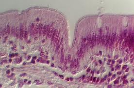
Histological section of intestine lining
Sticking with the immune system, many chronic diseases have inflammation as an underlying cause. Inflammation is an essential part of our immune response, but is only beneficial if it is transient; sustained inflammation leads to damage around the body. It has been found that Bifidobacteria and Lactobacilus species in the gut are essential for maintenance of the epithelial lining in this organ (the cells that make up the walls of our gut). A proper epithelial lining plays an essential role in the passage of nutrients out of our digestive system into our blood. The lining needs to be ‘selectively permeable’ so that only certain, useful, things get through. If the lining becomes ‘leaky’, then unwanted molecules can get through such as bacteria and their toxins or whole protein molecules (instead of just the amino acids we normally absorb from the gut), all of which could trigger an inappropriate immune response. Since Bifidobacteria and Lactobacilus are needed for maintenance of the epithelial lining, any disruption to these bacteria will have an impact on the integrity of the barrier. Indeed, it has been shown that mice fed on a "junk food" diet have a disruption of these bacteria and develop a ‘leaky epithelia’. This caused a low level systemic inflammatory response, which eventually causes metabolic diseases. It is therefore highly possible that many disease with inflammation as an underlying cause could originate from issues with out internal bacterial species.

The final thing I'd like to discuss is that the notion of thinking with your gut may hold more truth than you know. It has been found that microbes in the gut are important for the generation of neurotransmitter molecules such as serotonin and thus may be playing an important role in regulating mood. Furthermore, there is evidence suggesting a link between the gut microbiome and the hypothalamic-pituitary axis (HPA), a region of the brain that shows disruption during clinical depressive episodes. Mice bred to have no microbiome show an enhanced stress response that can be curtailed by the introduction of a microbiome - this response, to a large extent, is generated from the HPA. Additionally, it been shown in mice that if the microbiome of adventurous mice is transplanted into the guts of timid mice they lose their inhibitions and become more adventurous, further supporting the notion that our gut bacteria may be influencing our brains.

It may be scary to think that we are only 10% human with the remainder being made up by microbes, the majority of which are bacteria. What may be even scarier to consider is that these bacteria may have an influence on our most evolutionarily important organ, the brain. Bacteria could be controlling our response to stress; they could also potentially have even broader and larger effects on our behaviours and mood. Additionally to affecting our brains, there is increasing evidence that the gut microbiome plays essential roles in our immune system and may even play a huge role in obesity. Obesity is one of the biggest issues in the Western world and any methods to tackle it are rightly gaining much attention. Since we can alter our gut microbiome through the foods we eat and the drugs we take it stands to reason that with increased research in the area we may be able to shape our microbiomes to tackle disease. Since the microbiome seems to affect so many different aspects of our physiology, altering it will be fraught with potential pit-falls. But to look at it in a more positive light, we may be able to tackle a plethora of diseases in a very simple and affordable manner. Bacteria tend to get a bad reputation, but without them we probably wouldn’t be here.