Wednesday, 2 January 2013

We need some CRE-ative solutions


“Superbug” is an expression we hear far too often. Due to our excessive use of antibiotics over the past 65 or so years we have excelled this term from a potentially comical image of a flying inset that fights crime, to something we genuinely need to fear in a medical context. Admittedly, I am not a fan of the term “superbug,” but it is certainly catchier than the more accurate description of drug resistant or multi-drug resistant bacteria. The bacteria that will most likely spring to mind as being “superbugs” are MRSA and C. difficile. Both of these bacteria are well known to plague hospitals worldwide and cause potentially life-threatening disease in those people at their most vulnerable (in hospital for some other aliment). While MRSA and C. diff. are well established as health care-associated resistant bacteria, a new player is starting to come to the attention of healthcare officials and its emergence is being greeted with genuine fear.

Before discussing these new resistant bacteria I’d first like to talk about why we have resistant bacteria at all. In one sense, we can blame evolution. Bacteria are always looking to survive and will evolve to deal with anything that threatens this. The emergence of resistance is a wonderful example of the process of natural selection that drives Darwinian evolution. Let us consider a situation in which a patient has a bacterial infection. If this patient takes antibiotics these will kill the bacteria and aid the patient’s recovery. However, not all bacteria are created equally and when they multiply the progeny can carry mutations that set them slightly apart from their parent. It is possible that these mutations confer the bacteria with a slightly increased chance of survival against the antibiotics. If this occurs then this, potentially lone, bacteria will survive at the expense of all the others and will continue to grow (‘survival of the fittest’). All the competition for space and recourses has been removed due to the death of all other bacteria so this resistant bacterium will thrive. In the face of continued antibiotic treatment any new mutations that confers even greater protection will cause the same affect, continually selecting the bacterium that has the best protection from the affects of the antibiotic. The logical end point for all of this is that a bacterium will emerge that is completely protected from an antibiotic and it will thrive and spread.

MRSA - probably the best known "superbug"
While it is easy to blame evolution for the emergence of resistant bacteria, that is not the full story; we as humans need to take a fair share of the blame. Since the discovery of a process for the mass production of penicillin shortly after the Second World War, we have overused antibiotics. Antibiotics have been overused in the context of bacterial infections and most likely have been used to treat bacteria that are not susceptible to that drug (only certain antibiotics are effective against certain bacteria). However, more worryingly there are many cases of antibiotics being used to treat infections not caused by bacteria, such as the common cold, which is ridiculous since the drugs only work on bacteria (like using a cough sweet to treat a heart problem). The reason that overuse, and incorrect use, causes an issue is that we have billions of bacteria inside us; put these in an antibiotic context and you promote the emergence of resistance. Consider the fact that MRSA is a strain of the bacteria Staphylococcus aureus, a bacterial species that the vast majority of us have naturally in our lungs and on our skin, the only difference is MRSA has evolved resistance to the antibiotic methicillin.

MRSA and other resistant strains of bacteria are dangerous, however we do still have some “last line of defence” antibiotics that can be pulled out at the last minute to save the day like a cheesy action film. However, more and more cases of a new type of resistance are being seen, particularly in the USA, known as CRE. CRE stands for carbapenem resistant Enterobacteriaceae, with carbapenem being one of our few remaining “last line” antibiotics. Enterobacteriaceae are a bacterial family that include many well-known bacteria such as E. coli, Salmonella, Shigella and Y. pestis (which caused the Plague).  These are the well-known species in the Enterobacteriaceae family; however so far, most cases of CRE have been seen in Klebsiella and Enterobacter species that cause lung, intestinal and urinary tract diseases.

CRE bacteria have been spreading through the USA since their first detection about 5 years ago. Studies have suggested that these bacteria are endemic in major population centres such as New York, LA and Chicago (a report suggests that 3% of patients in intensive care units in Chicago carry CRE bacteria). There are also small pockets of CRE throughout other areas of the USA. So far CRE cases have been confined to health care facilities such as hospitals and care homes, as is the case with other “superbugs” such as MRSA. However, as with anything there is always the potential for spread outside of these facilities. The thought of spread is a worrying one when you consider the fact that CRE infections currently carry a reported 40% mortality rate, much higher than that of MRSA. If I was writing this blog for a newspaper this is where I would leave that statistic, 40% of people who get CRE die, everyone be scared and fearful! However, mortality rates are a pretty useless statistic as they rely on the ability to detect every single case of CRE to be accurate, yet chances are, only the most serious (and therefore life-threatening) cases are likely to be detected, skewing the data in favour of a higher death rate. When you also consider that until recently CRE has not garnered much attention, meaning no-one has really been looking for it, you get a further skewing of the data (only the most serious will be taken notice of). There is also difficulty in detecting CRE bacteria since carbapenems are not readily used, in the hope of preventing the emergence of resistance. This is not to say we shouldn’t consider these bacteria as inconsequential and we should (as we are) be treating them with a well-deserved level of respect.

The bottom line is that CRE bacteria are resistant to our last remaining antibiotic against this family of bacteria. All that we can do at this stage is prevent, the age-old techniques of rigorous hand washing, protective clothing and isolation are the main ways to protect from CRE infection. If someone becomes infected there is no effective treatment, with the exception of some old antibiotics that have been shelved for years due to their high toxicity (not necessarily something you’d want to take).

CRE bacteria are something we should be rightly fearful of, however they also bring to the forefront a gapping hole in our medical research. While we have some new and improved antibiotics in the pipeline, very few of these are though to be of any use against CRE, and there is very little incentive for the major drug companies to look for ways to tackle CRE. The problem lies in the fact that antibiotics just don’t make enough money. Why would a major drug company want to spend a fortunate making a drug that is used for maybe a couple of weeks, until the patient recovers, when they could spend their money making drugs to target chronic diseases that will need to be taken for the rest of a patients life. Not only is the treatment time short, but also resistance emerges so quickly that any drug produced could rapidly become obsolete, making the money spent useless. It sounds wrong to neglect people who could die from an infection on the basis of economics, but unfortunately, that is how it is, in the bluntest (and a slightly cynical) way.

I titled this blog post as the fact that we need some creative solutions, both for the play on the CRE bacteria naming and because, simply put, we do. There are ideas out there, such as the use of phage to kill bacteria. Phage are viruses that only infect bacteria so if we can find a way to use these then it may provided a good alternative to antibiotics. There is also work being conducted to look for new and untapped sources of antibiotics with the discovery of new fungi and bacteria (which produce antibiotics) in obscure locations such as caves and oceans, however, as I’ve alluded to, there is a shortage of funding in these area.

Process of conjugation, one of the main ways resistance spreads
One potentially promising area of research may lie in targeting the mechanism used by bacteria to spread resistance. Resistance is controlled by the production of protective proteins from genes. It is very common that these genes are located on mobile genetic element known as a plasmid (stick with it as I will clarify what that means). Like us, bacteria have a genome that contains all the instructions needed to produce a functional bacterial cell. However, in addition to the genome many bacteria can have addition chunks of DNA (plasmids). These extra units of DNA float around the cell, separate from the rest of the genome, and can be passed from one bacterium to another. Think of this like books, if the genome is a full instruction book for producing the bacteria then a plasmid, is like a page taken from another book. These plasmids (extra pages) can move between bacteria through a structure known as a pilus, in a process often termed bacterial sex (due to some obvious similarities – Google it). If we can find ways to block this process, correctly known as conjugation, then we may be able to stop the spread of genes that confer resistance and maintain the efficacy of our current stock of antibiotics. However, as it stands, this is just a hypothesis.


CRE bacteria are worrisome, they are resistant to pretty much all of our current stock of antibiotics and the number of cases are on the up. There is a reported death rate of around 40% for CRE infection, and while this may be slightly skewed, it is still clearly a dangerous situation we find ourselves in. We need new and innovative ways to tackle these bacteria as simply stopping the spread with preventative measures can only go so far. However, whether the funding will reach the research into these areas is yet to be seen. 

Friday, 30 November 2012

Restricted access post 3 of 3


I left the last blog touching on the fact that human schalfen 11 may potentially be able to act on influenza virus in addition to HIV. One protein that is already established as an influenza restriction factor is IFITM3, the topic of this blog post. The reason research in the field of IFITM3 is of much interest is that our current stock of drugs to tackle the virus are struggling. We currently have four licensed drugs to tackle influenza, however only two of these remain truly effective due to the emergence of resistant strains. Both Tamiflu and Relenza remain effective, but there are a growing number of cases of Tamiflu resistant influenza. If Tamiflu resistance continues to spread we will be left with only Relenza as an effective drug. Having a single drug is never a great situation as it makes it very likely that resistance will emerge, which would leave us with nothing! We therefore need novel approaches to tackle influenza; enter IFITM3…

IFITM3 was first defined in 2009. The protein was identified in a large-scale screen of proteins that are ‘turned on’ when flu infects cells. This switching on is controlled by a set of immune proteins known as interferons (IFN) that are produced by cells when they are infected (IFITM stands for InterFeron Induced TransMembrane protein). The IFN response turns on around 2000 protective proteins that produce an ‘antiviral state’ to help protect cells from the invading virus. The team wanted to find which of these 2000 or so proteins are most important for tackling influenza infection. In order to do this they systematically switched off individual proteins to see which ones provided the most protection (defined by exacerbated infection in the absence of the protein). By this approach they found 120 proteins that play an inhibitory role on influenza infection. Through further probing they found the best “hit” was the IFITM3 protein. IFITM3 is closely related to two other proteins, IFITM1 and IFITM2 (as the names would suggest), and both of these proteins were also found to inhibit influenza infection, albeit to a lesser extent.

The IFITM proteins play a major role in protecting cells from influenza based on the fact that their removal from cells allows a higher level of infection, as seen in the study published in 2009, and many other since. Interestingly it isn’t only influenza that these proteins protect us against. The team that originally identified the IFITM proteins found that they could also protect cells from West Nile and Dengue viruses (WNV and DENV). These two additional viruses are closely related to each other, but not very closely related to influenza, making the finding somewhat mysterious.  Since the original discovery, IFITM proteins have been found to act on yet more viruses including Marburg, SARS, the highly feared Ebola and HIV (though there is some debate about the link to HIV).

As with most things in biology, viruses are grouped together based on certain characteristics and their evolutionary origins, just like animals. As I have mentioned WNV and DENV are very closely related (let’s say similarly to chimps and humans) and are in the same viral evolutionary family, the Flaviviridae. The SARS virus is similar to the flaviviruses but is within a different family, known as the Coronaviridae (more distantly related, such as humans/chimps compared to orang-utans).  Influenza falls into a different viral family (Orthomyxoviridae) and is more distantly related to the flaviruses and coronaviruses (let’s say like a domestic cat compared to the apes). However influenza is reasonably closely related to the Filoviridae family that contains Ebola and Marburg viruses (like a house cat to a lion). HIV is not particularly related to any of the others mentioned here and falls into the family of Retroviridae (like a kangaroo to all the other mentioned animals). I direct your attention to the image to see how these families all interconnect. The viruses that the IFITM proteins target are all very diverse, which begs the question of how these proteins are able to target such a wide array of invaders. (I’d just like to point out that the animal examples I have used are just for demonstration).
Relationship of different viral families
 Even though the IFITM sensitive viruses are very diverse, they all, with the exception on HIV (hence the dispute), enter cells in a very similar manner. Initially the virus binds to receptors on the surface of a cell, somewhat akin to grabbing a door handle. Once bound the virus is able to enter into the cell (open the door) through a process known as endocytosis.  At this point the virus is essentially encased within a small bubble inside the cell that is known as a vesicle (think of a bubble within a glass of drink, sorry to mix metaphors). This vesicle joins up to a larger structure known as an endosome, which over time becomes acidic. This acidification eventually causes a change to the virus that allows it to fuse with the bubble and escape to the rest of the cell where it will then move on to complete its life cycle. Don’t be distracted by the names (or the metaphors), just have a look at the picture if my description has been a bit hard to follow. The main point to take from this paragraph is that all of the viruses that the IFITM proteins inhibit, except HIV, enter cells through this endocytosis and acidification process.

Entry of influenza through the endocytic pathway

Since all the viruses that IFITM has been found to have an inhibitory affect on enter cells in the same way it seems fair to assume that the IFITMs must be targeting this pathway. Evidence is stacking up that this is indeed the case as numerous studies have found that IFITM3 (less work has been done on the other two) is localised to vesicles and the endosome. However, it is still not clear exactly how the IFITM proteins actually interfere with the processes involved with the viral life cycle. The current hypothesis is that the protein may interfere with acidification, which would block exit of the virus from the endosome. This eventually leads to destruction of the virus since the acidification continues to a point that kills the virus. This idea is supported by the fact that less virus is seen inside the cell cytoplasm when IFITM3 is present (the cytoplasm is simply the contents of the cell, using the analogy of the vesicle being like a bubble in a drink from before; the cytoplasm is the liquid).

A comparison of the different entry mechanisms used by Influenza and HIV
We need to fully resolve how the IFITM proteins are functioning. Once that has been achieved it will be possible to look towards finding drugs that act in a similar fashion. If we are able achieve this, we may well be able to develop drugs that are highly active against a very broad set of viruses. In addition, these potential drugs would be targeting a human process meaning that resistance is less likely to evolve since humans mutate so very much slower than viruses. That’s a long way off yet but shows why we need basic research into these proteins.

As an extra little add-on, IFITM3 has been linked to severe flu in patients as well as simple studies in a lab. A study was conducted to look at patients hospitalised by pandemic H1N1 (Swine Flu) and season flu and found that these patients were enriched for a certain mutation to the IFITM3 gene (the mutation was more common in people who were hospitalised than the general public). This hints towards the fact that IFITM3 truly does impact on influenza and helps most people to control it (flu is rarely fatal). It also hints towards the fact that this mutation could potentially be used to find those people who are at the highest risk of influenza infection, which would help with deciding who receives the limited stocks of influenza vaccine produced each year.

IFITM3 is a very interesting protein that has the potential to make a large impact in our efforts to tackle influenza (and potentially other viruses). I will be keeping a close eye as the story develops and will, in all likelihood, write future blogs on the topic as the story evolves.

Monday, 19 November 2012

Restricted access (post 2 of 3)


Those who read my previous blog will have been introduced to viral restriction factors and will, hopefully, remember that my inspiration for writing on such a topic was the discovery of a new member of these proteins; human schlafen 11. Those who didn’t read the previous post may find it useful to go back so as to aid in the understanding of this post.  Besides the discovery of a new restriction factor, this paper caught my eye as a wonderful demonstration of how logical and step-by-step science can be. Hopefully I will be able to convey this logical progression through this blog post, as it really is a very nicely written paper.

As I mentioned in the previous post, the paper in question is from Michael David’s lab at University of California, San Diego and has now been published in theprint issue of the journal Nature, as of the 1st November.  The paper centres on the schlafen genes (SLFN), of which there are six in humans. These proteins are of particular interest since they have been consistently shown to be highly expressed in cells and organisms that are infected with viruses or bacteria, yet they had no known function. Along with the data from in vivo studies (in living organisms such as mice), the team also looked at cell lines (collections of cells grown in a lab) and found that there was differential expression between two very closely related cell lines, HEK293 and HEK293T cells. They found that HEK293 cells (hereafter referred to simply as 293 cells) express high levels of SLFN5 and SLFN11, while the HEK293T cells (293T cells) did not. Finding this different level of expression gives the team two cell lines that are easy to conduct experiments in (unlike animals) and allows for easy comparison of the situation with and without the SLFN proteins.

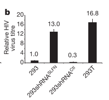
An example of their data. Virus titre is the level of released virus.
293 cells show low levels of release. The removal of SLFN genes from
these cells (by shRNA) causes increased release of virus. The 293T cells
show a high level of virus release.
The first experiment the team conducted was to look at the effect of removal of the SLFN genes from 293 cells (the cells that express) and compare this with the 293T cells and normal 293 cells. Any new similarities seen between the 293T cells and the 293 cells that have had the SLFN genes removed may be due to the lack of SLFN genes. If these similarities are different to normal 293 cells then it is even more likely to be due to the lack of SLFN genes (sorry if that is hard to follow with the similar nomenclature). The team infected the cells with multiple viruses including a HIV virus that causes cells to glow when it integrates into the genome (I refer you back to the previous post for details of the HIV life cycle if you are unfamiliar). They found that this HIV virus was able to integrate into all three of the different cell types. However, the cells that were lacking for SLFN11 released a much higher level of new virus that those that had the protein. This indicates that SLFN11 is applying some sort of restrictive affect on the HIV life cycle between integration and release of new virus from the cell. (The removal of SLFN5 had no affect so will not be mentioned again).


To only show that removal of SLFN11 allows more virus to be released does not give a full picture, so the team set out to further confirm the results that SLFN11 is applying a block to the HIV life cycle. To do this they went the other way, instead of removing the SLFN11 protein from cells that usually have it, they added it to cells that do not usually have it (the 293T cells). Addition of SLFN11 to 293T cells caused a substantial reduction in the level of virus released from the cells and this reduction was of a similar magnitude to that seen in the 293 cells that were naturally expressing the protein. This gives two lines of evidence that SLFN11 is indeed causing a block to the HIV life cycle after integration.

There are multiple steps between HIV DNA becoming integrated to the host and the release of viral particles. In the interest of developing a full understanding of the role SLFN11 is playing the team began to zoom in on exactly where the protein was having its restrictive affect. They took the logical approach of looking at the next step after integration. Once HIV DNA is integrated to the host genome it is used to make proteins via an intermediate of RNA. This is how all proteins are made; DNA acts as a template to make RNA that is then used as a code to make protein. The team looked to see if there was any difference in the level of HIV RNA produced in the cells that expressed SLFN11 compared to those that don’t. Unlike the results seen for release of new virus, the presence of SLFN11 had no affect on the levels of HIV RNA seen inside the cell. The window in which SLFN11 causes a block is narrowing, we now know the block occurs between production of RNA and release from the cell.

Having ruled out the first step post-integration the team moved on to look at the final step of the HIV life cycle; release from the cell. As I mentioned in the previous post, the protein tetherin acts at this final stage and causes accumulation of virus particles on the cell. The team looked for similar affects in the SLFN11 expressing cells and saw none. We now have evidence that the block is occurring between production of RNA and before the virus starts to leave the cell. This pretty much leaves two stages, production of proteins and assembly of these proteins into new virus particles.

Knowing that SLFN11 was either affecting protein expression or assembly of the virus the team looked for the presence of viral proteins inside the cells. They found that there was substantially less viral protein inside the cells that expressed SLFN11 even though there was the same amount of RNA from which the protein is made. The really interesting thing about this is that human proteins being expressed in the cells were completely unaffected. This means SLFN11 is causing a block to viral protein expression but not host protein expression; the protein is therefore highly specific to the invading pathogen. The team further confirmed this finding by adding artificial DNA to a cell that coded for a viral protein or a non-viral protein and saw that only the viral protein was blocked for expression. The evidence with both actual virus and with artificial DNA strongly supports the idea that SLFN11 is blocking viral proteins from being produced, and is doing so in a specific manner.

The question then becomes why. Why is it that SLFN11 only affects viral protein production? What is different between the RNA of the virus that makes protein compared to the human RNA that makes protein? For me to answer this question I need to tell you a small bit about DNA.  A molecule of DNA is made up of chemicals known as nucleotides of which there are four, known as A, T, C and G. In the DNA double helix, A always binds to T while C always binds to G. It has been observed that viruses and humans have different biases in the nucleotide composition of their genomes. Humans have a bias towards GC nucleotides (around 60%) in our DNA, while certain viruses have a bias towards AT nucleotides (I will however refer to the viruses as low GC, though this is obviously the same as high AT). 
An illustration of DNA. I have mentioned A, T, C and G which is the short hand
for the chemicals listed in this image.


Knowing of the difference in nucleotide bias between humans and viruses, the team asked whether SLFN11 was taking advantage of this. To address this question they produced two artificial DNA constructs for a HIV protein known as gag. One of these constructs used the normal sequence for gag (low GC content) while the other construct changed all the nucleotides that could be changed, without affecting the protein, to C or G, so to shift the bias towards that of a human gene (a gag gene that is high GC). They found that expression of the normal gag construct was inhibited, as expected, while expression of the high GC gag gene was not inhibited by SLFN11 at all. This gives good evidence that SLFN11 is indeed taking advantage of the differences between the nucleotides used by HIV compared to those used in humans.

A further validation of the findings was once again pursued. To achieve this the team took a protein known as enhanced green fluorescence protein (EGFP) that can be expressed in human cells and, as the name implies, fluoresces green. When the team changed the EGFP DNA to have low GC (so to make it like a HIV gene) it was seen that SLFN11 inhibited the levels of fluorescence in the cells, so is therefore inhibiting EGFP protein production. This evidence further supports the idea that the SLFN11 protein is specific to viral proteins as a function of their nucleotide bias, not some other function of their genome.

That concludes the story… sort of. It still isn’t clear exactly how SLFN11 blocks expression of proteins, but we know that it does, and we know why it is highly specific to viral proteins. Hopefully this post hasn’t been too hard to follow for any non-scientists reading, I’ve tried my best to explain without making the post overly long. I appreciate that I may have skipped over some parts particularly surrounding expression of proteins, so if anything is unclear please feel free to leave a comment and ask a question. I also hope I have managed to convey what I perceive as the beautifully logical progression of this paper. The team found that their protein did not stop HIV integration but did stop release of new viruses. They then looked to zoom in on exactly where this block was occurring by showing that RNA production and release of virus were not altered. This led them to look at the production of proteins where they found a substantial block in the presence of SLFN11. The question then became why was SLFN11 specifically blocking viral proteins but not human proteins. A question answered by looking at the different bias of human and viral DNA for G and C nucleotides. Hopefully it also shows how science relies on negative and positive confirmations of findings, for instance, when no SLFN11 then observe more virus release (negative), while the addition of SLFN11 to these cells causes a block (positive), giving very nice proof to the role of SLFN11.

Not only is the discovery of SLFN11 very interesting in regards to HIV but since it targets all genetic material that has a bias towards low GC content (as shown with the EGFP experiment) this implies it would be able to target other viruses. One such virus with a similar bias as HIV is influenza. If we can make drugs that act in a similar way we may well be able to make broadly acting anti-viral drugs that are highly specific to virally infected cells, helping to reduce their side affects and improve their efficacy.

The fact that SLFN11 may have the potential to act on influenza leads me nicely into the topic of my next post, the protein IFITM3. Make sure you come back for that if you’re interested.